
Tag Archives: ankle
Ankle Syndesmosis Anat Image
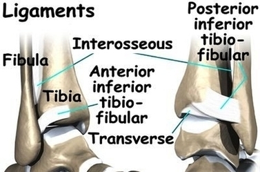
The ankle syndesmosis sits next to the ankle synovial joint, where the tibia meets the talus bone. The ankle syndesmosis is supported and held together by three main ligaments. Injuries to the syndesmotic ligaments of the ankle or “high ankle View Diagram Ankle Syndesmosis Anat Image
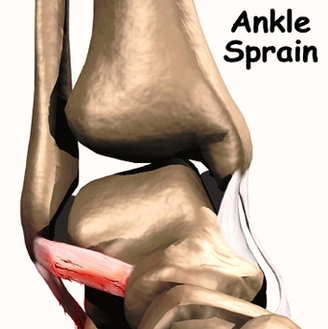
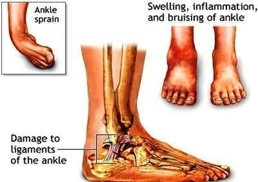
Ankle Sprain Causes Image
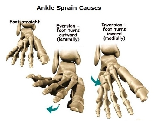
A sprain occurs when your ankle is forced to move out of its normal position, which can cause one or more of the ankle’s ligaments to stretch, partially tear or tear completely. Causes of a sprained ankle might include: Factors View Diagram Ankle Sprain Causes Image
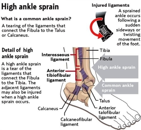
Diagram Of High Ankle Sprain Image
Diagnosing a high ankle sprain. Because syndesmotic sprains can be associated with lateral ligament injuries, medial ligament injuries, and fractures of the fibula, x-rays of the lower leg and ankle are necessary. If the athlete has a total syndesmosis rupture, View Diagram Diagram Of High Ankle Sprain Image
Sprained Ankle Rehab Image
You can do rehab exercises at home or even at the office to strengthen your ankle. How to do rehabilitation exercises for an ankle sprain How to do rehabilitation exercises for an ankle sprain Start each exercise slowly and use View Diagram Sprained Ankle Rehab Image
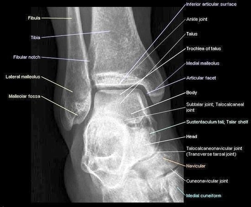
Foot Ankle Lateralview Diagram Image
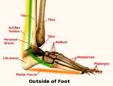
Ankle lateral view is part of a three view ankle series; this projection is used to assess the distal tibia and fibula, talus, navicular, cuboid, the base of the 5 th metatarsal and calcaneus. Article: Patient position. The anatomy of View Diagram Foot Ankle Lateralview Diagram Image
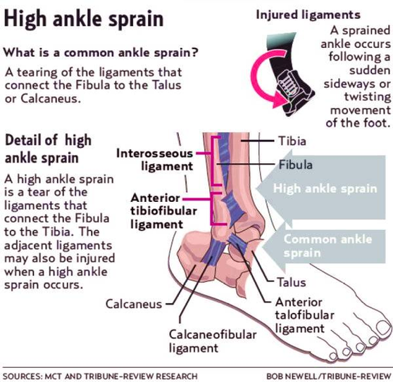
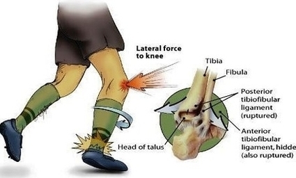
High Ankle Sprain Cause Diagram Image
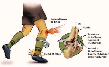
A high ankle sprain occurs when the ligaments above and around the ankle (see image below) get damaged or over-stretched. High ankle sprains are more serious than other ankle sprains because these specific ligaments hold the two lower bones of View Diagram High Ankle Sprain Cause Diagram Image
Ankle Sprains Diagram Image
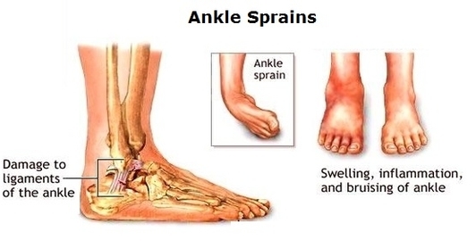
Bruising and swelling are common signs of a sprained ankle. If there is severe tearing of the ligaments, you might also hear or feel a “pop” when the sprain occurs. Most sprained ankles occur in the lateral ligaments on the View Diagram Ankle Sprains Diagram Image
Ankle Sprain Chart Image
A Grade I ankle sprain is the least severe. This is a stretching of one or more of the ligaments which results in mild pain and tenderness. Usually, the patient can bear weight and has only mild stiffness in the View Diagram Ankle Sprain Chart Image
Ankle Anterior View Radiograph Large Image
Radiographic analysis of the ankle often includes three views: the anteroposterior view, the internal oblique view, and the direct lateral view (15). The anteroposterior view helps assess the ankle mortise through the lateral portions of the talus and tibiotalar joint View Diagram Ankle Anterior View Radiograph Large Image
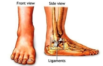
Sprained Ankle Anatomy Image
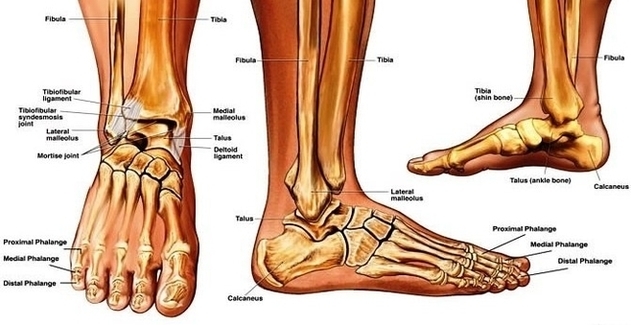
Ankle Anatomy1 Image
1,365 ankle anatomy stock photos and images available, or search for foot and ankle anatomy to find more great stock photos and pictures. The anatomy of the foot is incredibly complex. This introduction to the anatomy of the foot and View Diagram Ankle Anatomy1 Image
Ankle Joint Anatomy On Healthfavo Image
The ankle joint is an important joint in the human body, having a wide range of movements and consisting of different bones and ligaments. Learn now! This is an article covering the anatomy of the Tibia – interaction with the View Diagram Ankle Joint Anatomy On Healthfavo Image
Ankle Sprain Anat Image
A sprained ankle can occur on the lateral side of the ankle (most common), the medial side of the ankle (least common) or can occur as a syndesmotic sprain when the ligaments between the distal tibia and fibula are injured, View Diagram Ankle Sprain Anat Image
High Ankle Sprain Cause Image
A high ankle sprain occurs when the ligaments above and around the ankle (see image below) get damaged or over-stretched. High ankle sprains are more serious than other ankle sprains because these specific ligaments hold the two lower bones of View Diagram High Ankle Sprain Cause Image
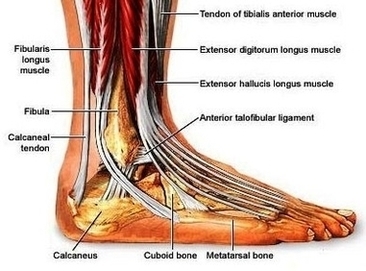
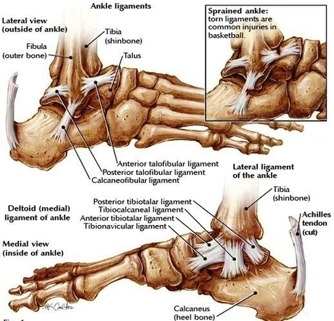
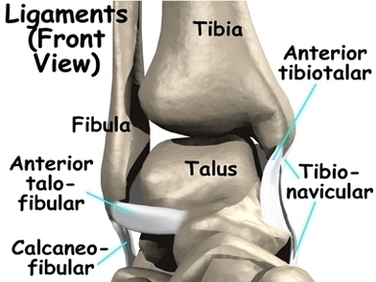
Ankle Ligaments Image
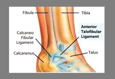
There are several major ligaments in the ankle: Three ligaments on the outside of the ankle that make up the lateral ligament complex, as follows: The anterior talofibular ligament (ATFL), which connects the front of the talus bone to the View Diagram Ankle Ligaments Image
Ankle Sprains Adam Image
A sprained ankle occurs when the ligaments are forced beyond their normal range of motion. Most sprained ankles involve injuries to the ligaments on the outer side of the ankle. Treatment for a sprained ankle depends on the severity of View Diagram Ankle Sprains Adam Image
Xray Normal Ankle Image
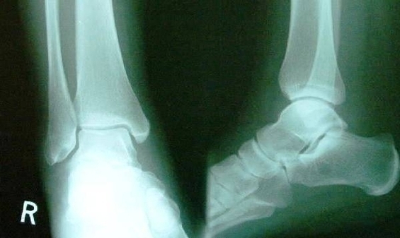
Interpreting an ankle X-ray. Use a methodical approach such as ABCs to look at a radiograph. Adequacy. Ideally, you should be able to see at least the distal third of the tibia and fibula and the talus on the mortise View Diagram Xray Normal Ankle Image
Treat An Ankle Sprain Step Image
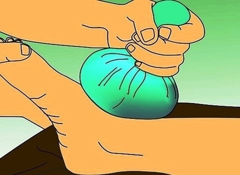
Read More… To treat a sprained ankle, apply an ice compress for 15-20 minutes every 2-3 hours until the swelling goes down. You can also manage swelling by keeping your ankle elevated for 2-3 hours every day and wearing a View Diagram Treat An Ankle Sprain Step Image
Ankle Sprains Types1 Image
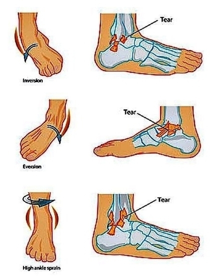
This is the most common type of ankle sprain and is characterized by moderate pain in addition to swelling, bruising, and some difficulty with movement. Third degree sprains occur when the ligament has torn completely. This is the most severe View Diagram Ankle Sprains Types1 Image