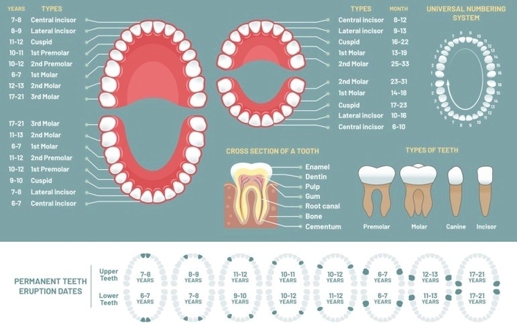
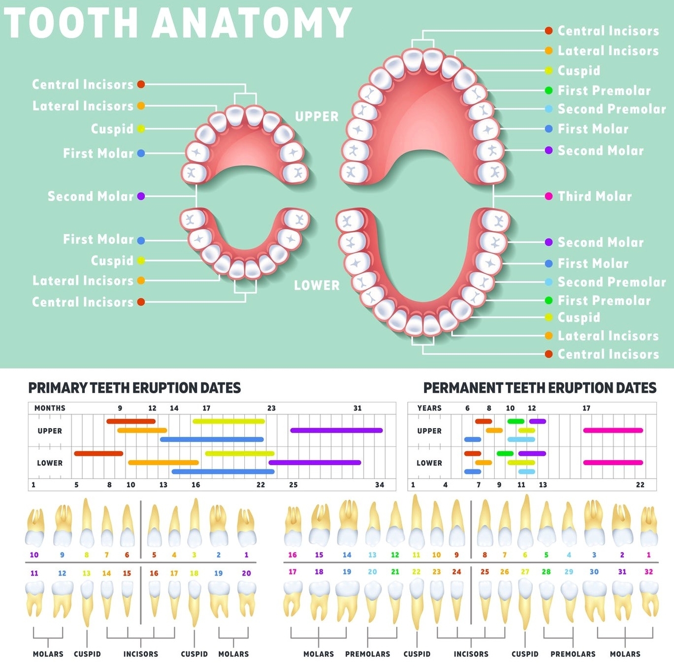
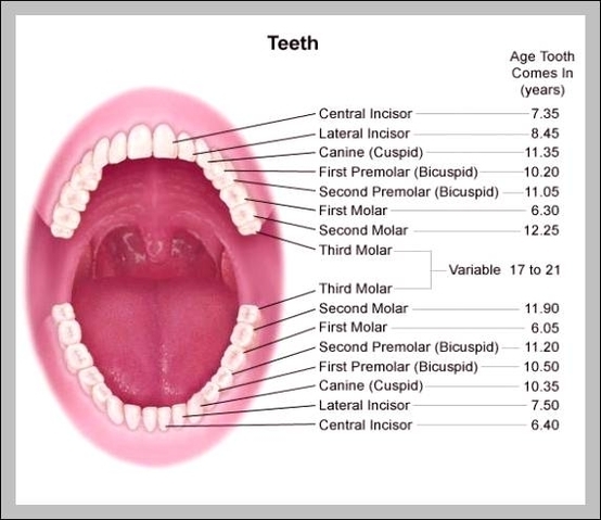
Permanent and Deciduous Teeth
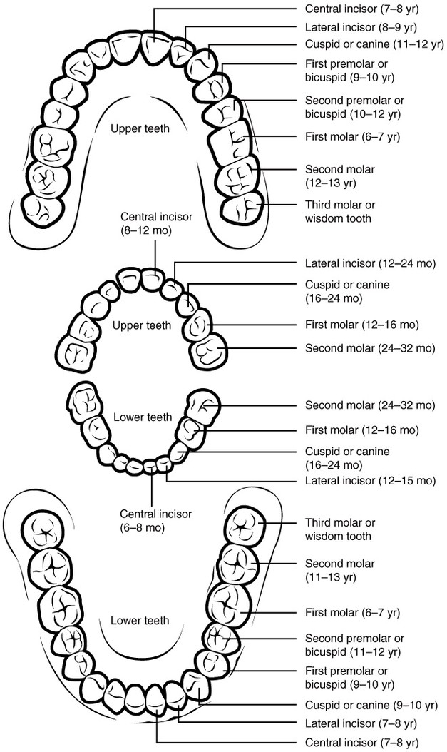
Permanent and Deciduous Teeth: Humans have two sets of teeth during their lifetime: deciduous (baby) teeth, which typically erupt between 6 months and 2 years, and permanent teeth, which replace them and are adapted for lifelong chewing and speech.