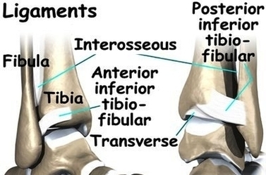
The ankle syndesmosis sits next to the ankle synovial joint, where the tibia meets the talus bone. The ankle syndesmosis is supported and held together by three main ligaments. Ankle Syndesmosis Anat Image Diagram - Chart - diagrams and charts with labels. This diagram depicts Ankle Syndesmosis Anat Image and explains the details of Ankle Syndesmosis Anat Image.
Ankle Syndesmosis Anat Image