Muscles attaching to the Pelvic Body

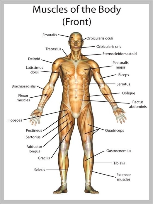
Muscles attaching to the pelvic body include the iliacus, psoas major, rectus abdominis, and pelvic floor muscles such as the levator ani. These muscles contribute to pelvic stability, posture, and locomotion, while also supporting pelvic organs. The iliopsoas muscles are View Diagram Muscles attaching to the Pelvic Body