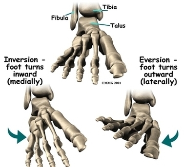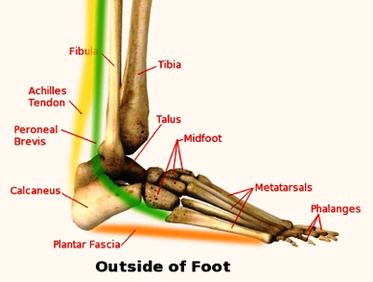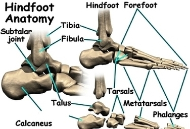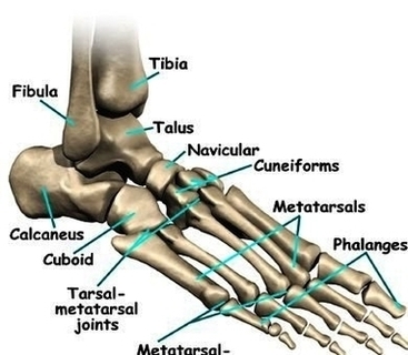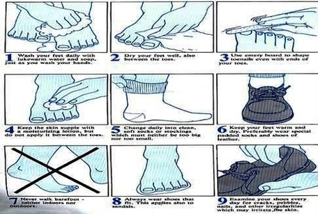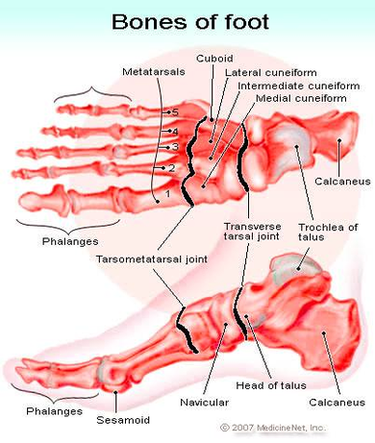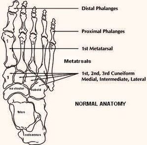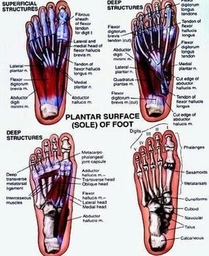
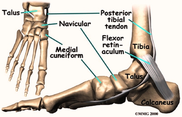
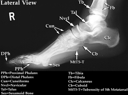

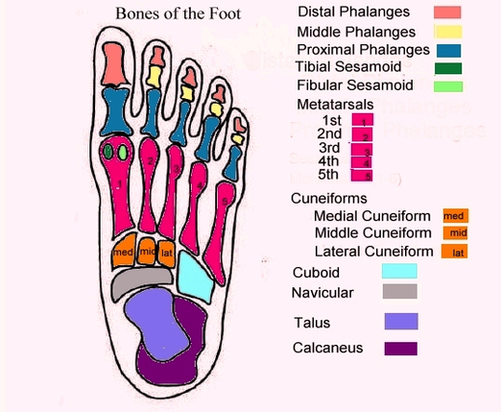
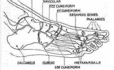
Foot Bones Diagram Image
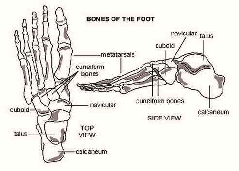
The skeletal structure of the foot is similar to that of the hand but, because the foot bears more weight, it is stronger but less movable. The bones of the foot are organized into the tarsal bones, metatarsal bones, and View Diagram Foot Bones Diagram Image