Category Archives: Health
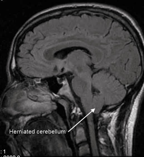
CT Scan of Cerebellar Tonsil Herniation
CT scans of cerebellar tonsil herniation show downward displacement through the foramen magnum, often associated with Chiari malformations. This can compress the brainstem, cerebellum, and cerebrospinal fluid pathways, causing headache, dizziness, motor or sensory deficits, and hydrocephalus. Imaging demonstrates the View Diagram CT Scan of Cerebellar Tonsil Herniation
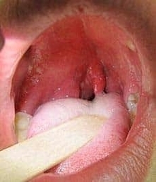
Quinsy Inflammation of Peritonsillar Tissue
Quinsy, also known as a peritonsillar abscess, is a collection of pus that forms in the tissue around the tonsil. It usually develops as a complication of untreated or severe tonsillitis. Patients often experience intense throat pain on one side, View Diagram Quinsy Inflammation of Peritonsillar Tissue
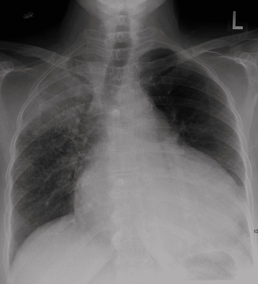
Pericardial Tamponade
Pericardial tamponade occurs when fluid accumulates in the pericardial sac, compressing the heart and impairing cardiac output. Knowledge of pericardial anatomy is essential for cardiologists, emergency physicians, and medical students in diagnosis, echocardiography, and pericardiocentesis. Understanding tamponade pathophysiology ensures timely View Diagram Pericardial Tamponade
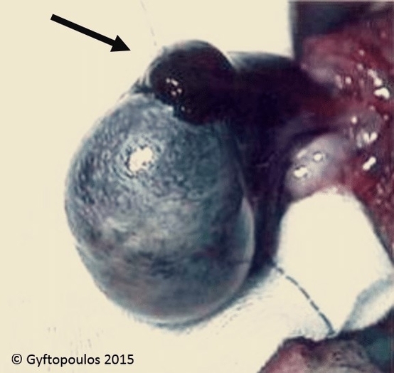
Testicular Torsion
Testicular torsion occurs when the spermatic cord twists, cutting off blood supply to the testis. It typically presents with sudden, severe scrotal pain and swelling. The affected testis may lie higher than normal and be tender to touch. Because prolonged View Diagram Testicular Torsion
Severe herniation of L4 L5 disc
A severe L4L5 disc herniation occurs when the nucleus pulposus protrudes through the annulus fibrosus, compressing nearby nerve roots. At this level, the L5 nerve root is commonly affected. Patients may experience lower back pain radiating down the leg, often View Diagram Severe herniation of L4 L5 disc
Episiotomy
Episiotomy is a surgical incision of the perineum during childbirth to enlarge the vaginal opening and prevent tearing. Knowledge of perineal anatomy is essential for obstetricians, midwives, and medical students in performing episiotomy, managing trauma, and postpartum repair. Understanding the View Diagram Episiotomy
Head Rotation
Head rotation mainly occurs at the atlantoaxial joint between the first and second cervical vertebrae. The dens of the axis acts as a pivot, allowing the atlas and skull to rotate around it. Muscles such as sternocleidomastoid and splenius capitis View Diagram Head Rotation
Mastoiditis Infection of the Mastoid Air Cells of the Ear
Mastoiditis is an infection of the mastoid air cells located in the mastoid process of the temporal bone. These air-filled spaces communicate with the middle ear, so infections often arise secondary to otitis media. The condition can cause swelling, pain View Diagram Mastoiditis Infection of the Mastoid Air Cells of the Ear
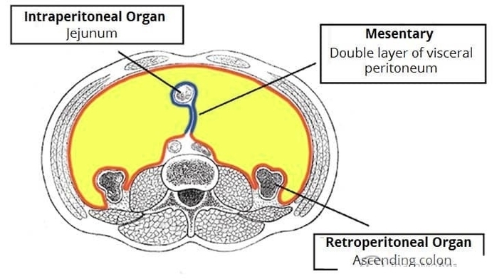
Intraperitoneal and Retroperitoneal Organs
Intraperitoneal organs, such as the stomach and small intestine, are completely enveloped by visceral peritoneum, whereas retroperitoneal organs, including the kidneys and pancreas, lie behind it. Understanding the distinction is critical for surgeons, radiologists, and medical students in planning surgical View Diagram Intraperitoneal and Retroperitoneal Organs
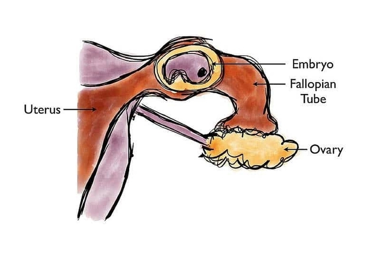
Ectopic Pregnancy
Ectopic pregnancy occurs when a fertilized egg implants outside the uterine cavity, most commonly in the uterine tube. As the embryo grows, it can cause stretching and eventual rupture of the tube, leading to severe internal bleeding. Patients often present View Diagram Ectopic Pregnancy
Positive Trendlenburg Test
A positive Trendelenburg test indicates weakness of the hip abductor muscles, particularly the gluteus medius, causing pelvic drop on the unsupported side during single-leg stance. Knowledge of hip mechanics is essential for orthopedic surgeons, physiotherapists, and medical students in diagnosing View Diagram Positive Trendlenburg Test
Septation of the Heart Embryology of the Cardiovascular System
Heart septation during embryology involves the formation of interatrial and interventricular septa, creating separate right and left chambers for efficient circulation. Knowledge of cardiac development is essential for pediatric cardiologists, surgeons, and medical students in managing congenital heart defects. Understanding View Diagram Septation of the Heart Embryology of the Cardiovascular System
Erbs Palsy Waiters Tip Position
Erbs palsy, often resulting from obstetric brachial plexus injury, causes a waiters tip posture. The affected upper limb is adducted, internally rotated, and the elbow extended due to paralysis of the shoulder and arm muscles innervated by C5C6 roots. Weakness View Diagram Erbs Palsy Waiters Tip Position
Aortic Aneurysm
Pacemaker Illustration Conducting System of the Heart
Pacemaker illustrations depict the hearts conduction system, including the sinoatrial node, atrioventricular node, bundle of His, and Purkinje fibers. The pacemaker delivers electrical impulses to maintain regular heart rhythm in patients with bradycardia, heart block, or arrhythmias. Placement requires understanding View Diagram Pacemaker Illustration Conducting System of the Heart
Varicose veins
Varicose veins are dilated, tortuous superficial veins, most commonly in the lower limbs, due to valve incompetence and venous hypertension. They can cause pain, swelling, and skin changes. Understanding the venous system, including the great and small saphenous veins and View Diagram Varicose veins
The Stages of Walking
Walking involves a complex sequence of alternating stance and swing phases coordinated by muscles, tendons, and the nervous system. Hip flexors, extensors, quadriceps, hamstrings, and intrinsic foot muscles control limb movement, balance, and propulsion. Understanding gait is essential in physiotherapy, View Diagram The Stages of Walking
Types of Spina Bifida
X Ray Generation Imagine
X-ray generation involves producing electromagnetic radiation through acceleration of electrons onto a target anode, creating photons that penetrate tissues and create radiographic images. Knowledge of X-ray physics is essential for radiologists, technicians, and medical students in imaging diagnostics, safety protocols, View Diagram X Ray Generation Imagine