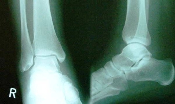
Xray Normal Ankle Image

This human anatomy diagram with labels depicts and explains the details and or parts of the Xray Normal Ankle Image. Human anatomy diagrams and charts show internal organs, body systems, cells, conditions, sickness and symptoms information and/or tips to ensure one lives in good health.

Interpreting an ankle X-ray. Use a methodical approach such as ABCs to look at a radiograph. Adequacy. Ideally, you should be able to see at least the distal third of the tibia and fibula and the talus on the mortise view and in addition to those, you should be able to see the calcaneum and the base of the 5 th metatarsal on the lateral view.

Use a methodical approach such as ABCs to look at a radiograph. Ideally, you should be able to see at least the distal third of the tibia and fibula and the talus on the mortise view and in addition to those, you should be able to see the calcaneum and the base of the 5th metatarsal on the lateral view.

Lateral view of the ankle tracing the outline of the tibia (green), fibula (red), talus (dark red), calcaneum (blue) and base of 5th metatarsal (orange). Note the os trigonum (red circle) which is an accessory ossicle of the foot and a normal variant. 3 Assess the joint space on the mortise view.

Xray Normal Ankle Image

Xray Normal Ankle Image


Tags: , ,