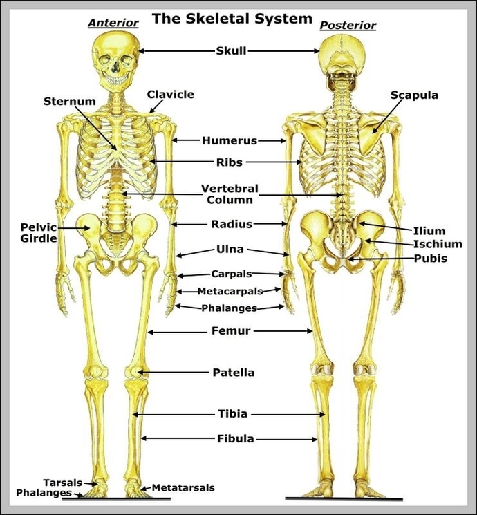
Skeletal System Pelvis Image

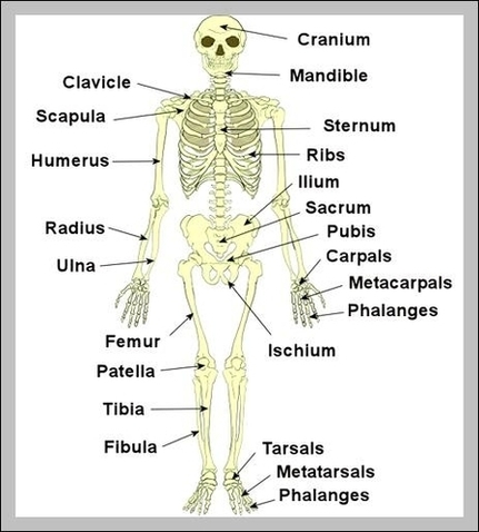
Human Pelvis Image Human male anatomy scheme. Main pelvic bones – sacrum, ilium, coccyx, pubis, ischium. Vector illustration isolated on a white background. pelvis anatomy stock illustrations Human male anatomy scheme. Main pelvic bones – sacrum, ilium, coccyx, pubis, ischium. View Diagram Skeletal System Pelvis Image