Anatomy Nose Large Image Diagram - Chart - diagrams and charts with labels. This diagram depicts Anatomy Nose Large Image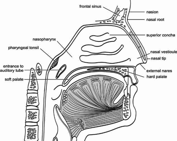
Tag Archives: large
Diagram Of Bronchial Asthmafig Large Image
The bronchi are lined with the same type of mucus that lines the rest of the respiratory tract. Foreign objects breathed into the lungs often end up in the right bronchus, as it is larger than the left. Once inside the lungs, each bronchus is further divided into five smaller, secondary bronchi, which provide air to the lobes of the lungs.
Once inside the lungs, each bronchus is further divided into five smaller, secondary bronchi, which provide air to the lobes of the lungs. The secondary bronchi continue to branch off to form the tertiary bronchi, which are further divided into terminal bronchioles.
They lead to the alveoli (tiny air sacs where the exchange of carbon dioxide and oxygen occurs) by way of alveolar ducts. Together, the trachea and the two primary bronchi are referred to as the bronchial tree. At the end of the bronchial tree lie the alveolar ducts, the alveolar sacs, and finally the alveoli.
Diagram Of Bronchial Asthmafig Large Image Diagram - Chart - diagrams and charts with labels. This diagram depicts Diagram Of Bronchial Asthmafig Large Image
Brain Anatomy Shirt Large Image
Brain Anatomy Shirt Large Image Diagram - Chart - diagrams and charts with labels. This diagram depicts Brain Anatomy Shirt Large Image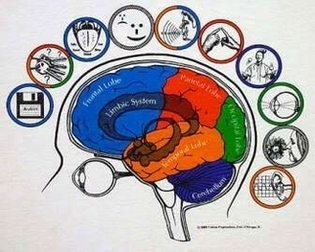
Ankle Anterior View Radiograph Large Image
Radiographic analysis of the ankle often includes three views: the anteroposterior view, the internal oblique view, and the direct lateral view (15). The anteroposterior view helps assess the ankle mortise through the lateral portions of the talus and tibiotalar joint overlap with the lateral malleolus (16).
A P view Lateral view Internal oblique view Three views of the ankle are mandatory for the evaluation of the ankle following trauma. Some fractures of the medial malleolus may not be apparent on the AP and lateral views but are readily visible on the internal oblique projection.
The AP view (Figure 11-1 A) shows the ankle mortise; however, the lateral margin of the talus and lateral portions of the joint are obscured by the underlying lateral malleolus. Soft tissue swelling seen about the medial and lateral malleolus serves as a useful clue to the presence of an underlying acute fracture. FIGURE 11-1
Ankle Anterior View Radiograph Large Image Diagram - Chart - diagrams and charts with labels. This diagram depicts Ankle Anterior View Radiograph Large Image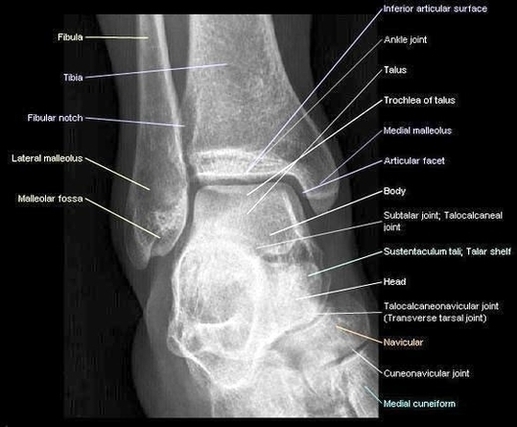
Human Body Large Image
399,556 the human body stock photos and images available, or search for the human body anatomy body or the human body photos to find more great stock photos and pictures.
Abstract image of a human body in the form of a starry sky or space, consisting of points, lines, and shapes in the form of planets, stars and the universe. Low poly vector background. the human body stock illustrations
internal organs and circulatory system Vector isolated illustration of human internal organs and circulatory system in man body. Stomach, liver, bladder, lung, kidney, heart, icon. Medical poster the human body stock illustrations Vector isolated illustration of human internal organs and circulatory system in man body.
Human Body Large Image Diagram - Chart - diagrams and charts with labels. This diagram depicts Human Body Large Image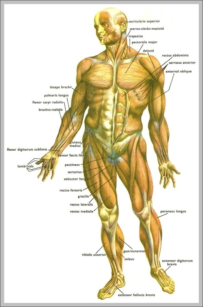
Teeth Anatomy Large Image
Key facts about tooth anatomy Types of teeth Deciduous dentition (20 teeth), permanen … Teeth Numbering/notation systems ISO/FDI system (International standard), … Main parts of a tooth Crown, pulp chamber, neck, dental root, … Neurovascular supply Arteries: anterior superior alveolar art …
It is a hard tissue that contains microscopic tubes. When the enamel is damaged, heat or cold can enter the tooth through these paths and cause sensitivity or pain. • Pulp: The softer, living inner structure of teeth. Blood vessels and nerves run through the pulp of the teeth.
318,995 human teeth stock photos, vectors, and illustrations are available royalty-free.
Teeth Anatomy Large Image Diagram - Chart - diagrams and charts with labels. This diagram depicts Teeth Anatomy Large Image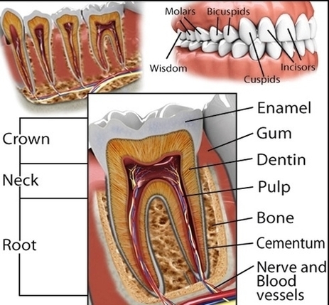
Cranial Nerves Anatomy Brainstem Human Body En Large Photo Image
Cranial Nerves Anatomy Brainstem Human Body En Large Photo Image Diagram - Chart - diagrams and charts with labels. This diagram depicts Cranial Nerves Anatomy Brainstem Human Body En Large Photo Image
Large Tooth Diagram Image
4,030 teeth diagram stock photos, vectors, and illustrations are available royalty-free. See teeth diagram stock video clips
A tooth chart consists of illustrated images of each tooth in the mouth. Not only images, but each tooth also has a unique name and shape to be identified. In tooth charts, there are different types of descriptions.
When looking at a human teeth diagram, it’s important to remember perspective. The diagram is set up to view the teeth from the perspective of the dentist. What is seen on the left side of the diagram is actually the right side of the patient’s mouth. The teeth numbers start on the patient’s right.
Large Tooth Diagram Image Diagram - Chart - diagrams and charts with labels. This diagram depicts Large Tooth Diagram Image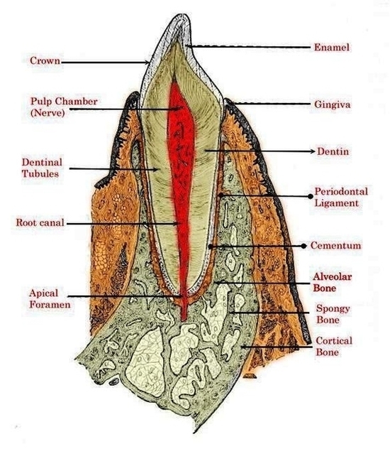
Forming Tumour Large Image
Drawing shows different sizes of a tumor in centimeters (cm) compared to the size of a pea (1 cm), a peanut (2 cm), a grape (3 cm), a walnut (4 cm), a lime (5 cm), an egg (6 cm), a peach (7 cm), and a grapefruit (10 cm). Also shown is a 10-cm ruler and a 4-inch ruler. Tumor sizes are often measured in centimeters (cm) or inches.
10,589 brain tumour stock photos and images available, or start a new search to explore more stock photos and images. x-ray of brain showing tumor x-ray of brain showing tumor. Computer generated images with correct male anatomy, showing brain with a glowing tumor inside. brain tumour stock pictures, royalty-free photos & images
208,642 tumor stock photos and images available, or search for tumor cell or cancer cells to find more great stock photos and pictures.
Forming Tumour Large Image Diagram - Chart - diagrams and charts with labels. This diagram depicts Forming Tumour Large Image
Diagram Heart Attack Large Image
A heart attack happens when your heart cannot get enough oxygen because the blood flow is abruptly disrupted. The heart muscle cannot pump properly, and tissue rapidly begins to die. [1] About 735,000 Americans have a heart attack every year.
The diagram of heart is beneficial for Class 10 and 12 and is frequently asked in the examinations. A detailed explanation of the heart along with a well-labelled diagram is given for reference. The upper two chambers of the heart are called auricles.
Pay attention to chest pain. A pain in the chest, whether it’s sharp or dull, is the most common sign of a heart attack. People who are having heart attacks often say they feel squeezing, fullness, pressure, tightness, or a sharp sensation in the center or left area of the chest.
Diagram Heart Attack Large Image Diagram - Chart - diagrams and charts with labels. This diagram depicts Diagram Heart Attack Large Image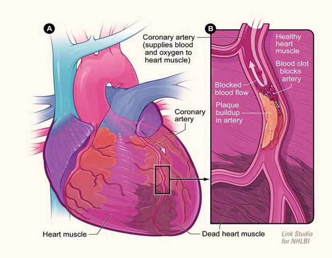
Diagram Of Human Anatomy Mediastinum Left View Illustration Large Image
Nerves: details the nervous anatomy of the mediastinum including notably the route of the phrenic and vagus nerves as well as the anatomy of the sympathetic trunk at the thoracic level. Heart: the external morphology of the heart in the mediastinum and the various chambers of the heart on the anatomical sections are detailed on these diagrams.
The superior mediastinum contains three visceral organs including the esophagus, the trachea and remnants of the thymus. The nerves that run through this area are three and there are also two different nervous plexuses.
Middle Inferior Mediastinum. The middle inferior mediastinum contains a single nerve which is the phrenic nerve, the heart and the pericardium. The vessels present include: the ascending aorta. the pulmonary trunk. the superior vena cava. the pericardiacoophrenic artery.
Diagram Of Human Anatomy Mediastinum Left View Illustration Large Image Diagram - Chart - diagrams and charts with labels. This diagram depicts Diagram Of Human Anatomy Mediastinum Left View Illustration Large Image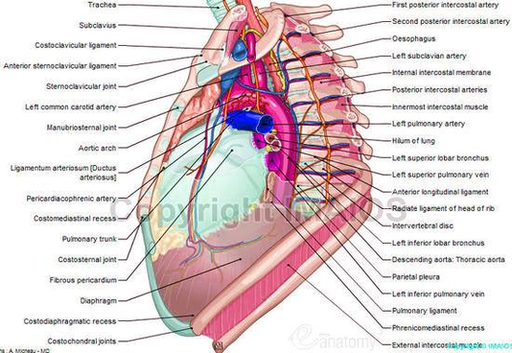
Nci Digestive Torso Large Image
Looking at pictures of your GI tract can help you to pinpoint where symptoms such as abdominal pain may be coming from. This understanding can also help you to better describe your symptoms to your healthcare provider.
The organs that make up the GI tract are the mouth, throat, esophagus, stomach, small intestine, large intestine, rectum, and anus. The GI tract is one part of the digestive system. 2
As you can see in the picture, intestinal contents move through the ascending colon, across the transverse colon and down through the descending colon. As material moves through the various parts of the large intestine, water and salt are absorbed by the lining and the material is compacted into the stool.
Nci Digestive Torso Large Image Diagram - Chart - diagrams and charts with labels. This diagram depicts Nci Digestive Torso Large Image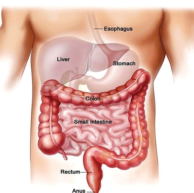
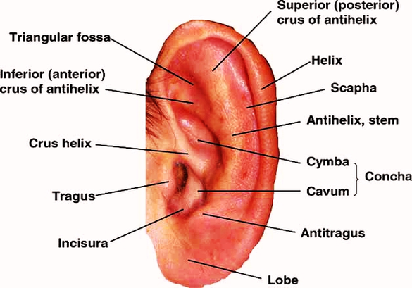
Anatomy Ear Large Image
Picture of the Ear. The spiral-shaped cochlea is part of the inner ear; it transforms sound into nerve impulses that travel to the brain. The fluid-filled semicircular canals (labyrinth) attach to the cochlea and nerves in the inner ear. They send information on balance and head position to the brain.
12,612 human ear anatomy stock photos, vectors, and illustrations are available royalty-free.
External Ear Anatomy (Auricle or Pinna) The outer ear auricle or external ear is composed of all of the parts of the ear outside the skull. It is also sometimes referred to as the auricle or the pinna. Although the outer ear is the least important part of the ear’s hearing function, it provides the necessary structure and protection.
Anatomy Ear Large Image Diagram - Chart - diagrams and charts with labels. This diagram depicts Anatomy Ear Large Image