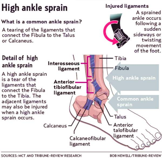
Diagnosing a high ankle sprain. Because syndesmotic sprains can be associated with lateral ligament injuries, medial ligament injuries, and fractures of the fibula, x-rays of the lower leg and ankle are necessary. If the athlete has a total syndesmosis rupture, separation will be evident in the x-ray between the tibia, fibula, and talus. Diagram Of High Ankle Sprain Image Diagram - Chart - diagrams and charts with labels. This diagram depicts Diagram Of High Ankle Sprain Image and explains the details of Diagram Of High Ankle Sprain Image.
Diagram Of High Ankle Sprain Image