Lateral Compartment of the Leg
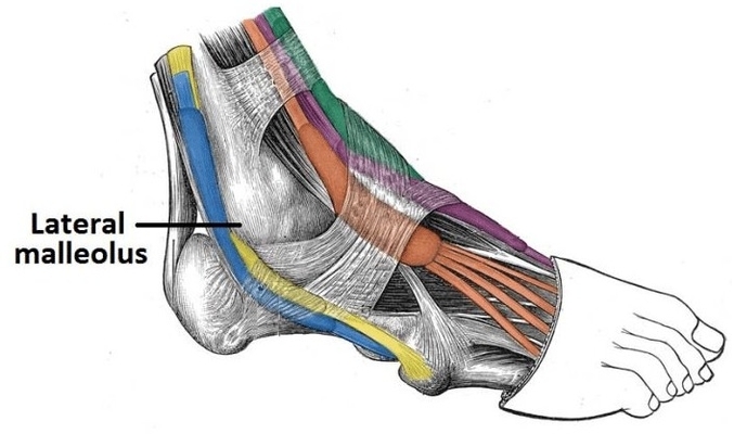
The lateral compartment of the leg contains the fibularis longus and brevis muscles, responsible for eversion and plantarflexion of the foot. These muscles are innervated by the superficial fibular nerve and receive blood supply from branches of the fibular artery. View Diagram Lateral Compartment of the Leg