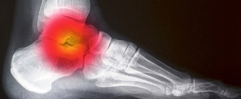

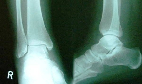
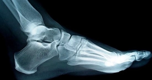
Foot Normal Lat Xray Image
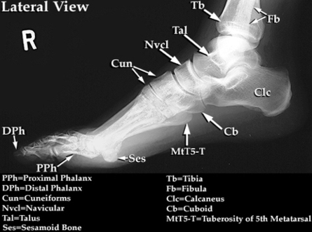
A normal foot x-ray will show three images of the foot, one from the front, one from the side, and one from the angle. The gamma ring is the smallest gamma ray and is not absorbed by all tissues. The View Diagram Foot Normal Lat Xray Image