Category Archives: Body Parts
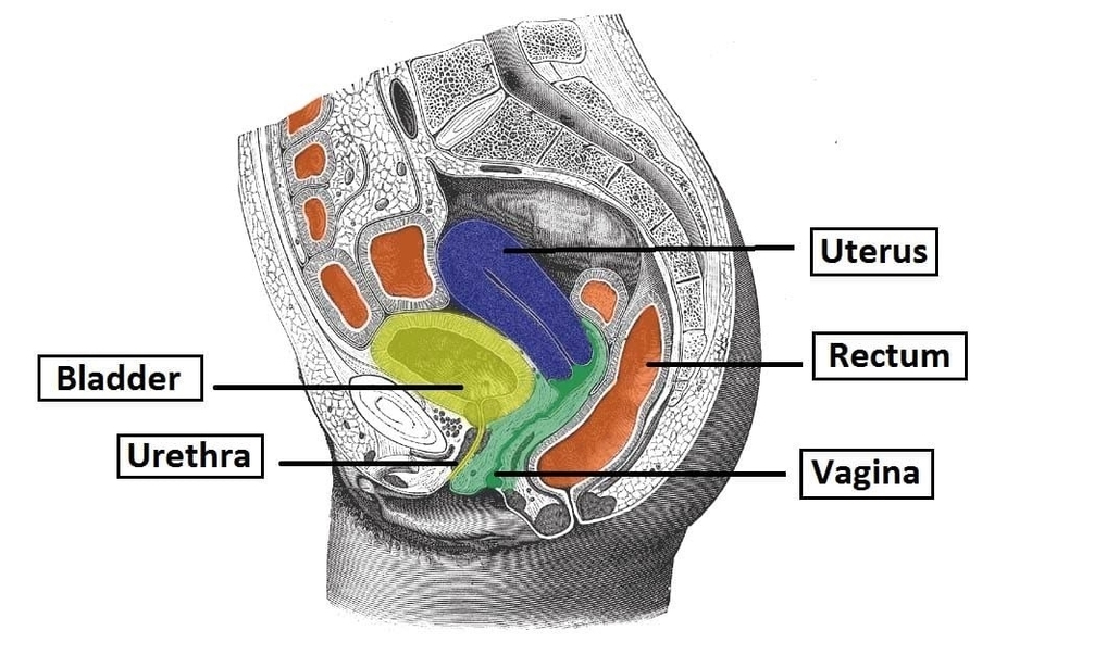
Overview of the Perineum
The perineum is the anatomical region between the pubic symphysis, coccyx, and ischial tuberosities, divided into urogenital and anal triangles. It contains muscles, fascia, neurovascular structures, and openings for the urethra, vagina, and anus. Understanding perineal anatomy is crucial in View Diagram Overview of the Perineum
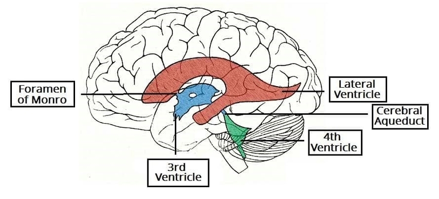
Superior View of Cast of Ventricular System
The ventricular system includes paired lateral ventricles, the third ventricle, cerebral aqueduct, and fourth ventricle, forming a pathway for cerebrospinal fluid circulation. Superior views of ventricular casts illustrate the precise arrangement of cavities and foramina. This system cushions the brain, View Diagram Superior View of Cast of Ventricular System
Anatomical Course of the Spermatic Cord
The spermatic cord is a vital structure of the male reproductive system that suspends the testis within the scrotum. This anatomy diagram of the anatomical course of the spermatic cord shows its formation at the deep inguinal ring, passage through View Diagram Anatomical Course of the Spermatic Cord
Contents of the Bony Orbit Arteries Optic Nerve and Muscles
The bony orbit houses the globe, extraocular muscles, optic nerve, and orbital vasculature. Arteries such as the ophthalmic artery supply the eye and surrounding structures, while cranial nerves III, IV, V1, and VI control movement and sensation. Ligaments, connective tissue, View Diagram Contents of the Bony Orbit Arteries Optic Nerve and Muscles
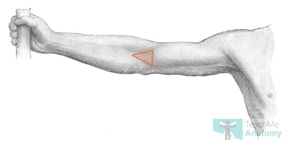
Anterior View of the Forearm Borders of the Cubital Fossa
The anterior view of the borders of the cubital fossa demonstrates its triangular boundaries formed by the brachioradialis and pronator teres muscles. This anatomy diagram highlights surface landmarks and underlying structures. It provides a clear understanding of the region used View Diagram Anterior View of the Forearm Borders of the Cubital Fossa
Prepubic and Infrapubic Angles of the Male Urethra
The prepubic and infrapubic angles of the male urethra refer to anatomical curves along the urethra that influence urinary flow and catheterization. Knowledge of these angles is essential for urologists, surgeons, and medical students in performing urethral instrumentation, managing strictures, View Diagram Prepubic and Infrapubic Angles of the Male Urethra
Distal End of the Spinal Cord The Lumbar Cistern
The distal end of the spinal cord includes the conus medullaris and lumbar cistern, containing cerebrospinal fluid and nerve roots of the cauda equina. The lumbar cistern extends from the conus to the sacral region, providing a safe site for View Diagram Distal End of the Spinal Cord The Lumbar Cistern
Suboccipital Triangle Borders and Vertebral Artery
The suboccipital triangle, located deep in the posterior neck, is bordered by the rectus capitis posterior major, obliquus capitis superior, and obliquus capitis inferior muscles. The vertebral artery and suboccipital nerve pass through this space. Knowledge of its borders and View Diagram Suboccipital Triangle Borders and Vertebral Artery
Medullary Pyramids
The medullary pyramids are paired structures on the ventral surface of the medulla oblongata, containing corticospinal tracts responsible for voluntary motor control. Knowledge of pyramidal anatomy is essential for neurologists, neurosurgeons, and medical students in managing strokes, lesions, or neurodegenerative View Diagram Medullary Pyramids
Anatomy of the Paranasal Sinuses
The paranasal sinuses are air-filled cavities within the skull that reduce weight and enhance voice resonance. This anatomy diagram of the anatomy of the paranasal sinuses illustrates the frontal, maxillary, ethmoidal, and sphenoidal sinuses. It highlights their drainage pathways into View Diagram Anatomy of the Paranasal Sinuses
Layers of the Scalp
The scalp consists of five layers remembered by the mnemonic SCALP: skin, connective tissue, aponeurosis, loose areolar tissue, and pericranium. The dense connective tissue layer contains blood vessels and nerves. The loose areolar layer allows movement of the scalp over View Diagram Layers of the Scalp
Cutaneous Innervation of the Radial Nerve Upper Limb
The radial nerve provides cutaneous innervation to the posterior arm, forearm, and hand, including the dorsal aspect of the lateral three and a half fingers. Knowledge of its distribution is essential for neurologists, orthopedic surgeons, and medical students in diagnosing View Diagram Cutaneous Innervation of the Radial Nerve Upper Limb
Contents of the Tarsal Tunnel
The tarsal tunnel is located on the medial side of the ankle, beneath the flexor retinaculum. It contains the tendons of tibialis posterior, flexor digitorum longus, posterior tibial artery, tibial nerve, and flexor hallucis longus. Compression of the tibial nerve View Diagram Contents of the Tarsal Tunnel
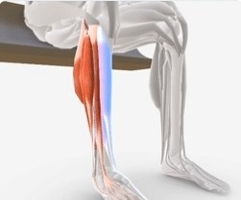
Ankle Dorsi Plantar
Dorsiflexion and plantarflexion are primary movements of the ankle joint that allow the foot to move upward and downward. This anatomy diagram of ankle dorsi and plantar movements illustrates how the talocrural joint enables controlled motion between the tibia, fibula, View Diagram Ankle Dorsi Plantar
Submental Triangle of the Neck
The submental triangle is a small triangular region located beneath the chin in the anterior neck. It is bounded by the anterior bellies of the digastric muscles and the body of the hyoid bone. The floor is formed by the View Diagram Submental Triangle of the Neck
Direct Inguinal Hernia vs Indirect Inguinal Hernia Regional Anatomy
Direct and indirect inguinal hernias are distinguished by their anatomical relationship to the inferior epigastric vessels. A direct inguinal hernia protrudes medial to these vessels through a weakened area of the posterior wall of the inguinal canal, often in older View Diagram Direct Inguinal Hernia vs Indirect Inguinal Hernia Regional Anatomy
Overview of the Mesentery
The mesentery is a double layer of peritoneum that suspends the intestines from the posterior abdominal wall, providing a pathway for blood vessels, nerves, and lymphatics. It anchors the small and large intestines while allowing for mobility during digestion. The View Diagram Overview of the Mesentery
Great Vessels of the Heart
The great vessels of the heart include the aorta, superior and inferior vena cava, pulmonary arteries, and pulmonary veins. These vessels transport blood to and from the heart, maintaining systemic and pulmonary circulation. Knowledge of their anatomy is essential for View Diagram Great Vessels of the Heart
Palmar Fascia of the Hand
The palmar fascia of the hand is a dense layer of connective tissue that invests the palm and fingers, forming structures like the palmar aponeurosis. It anchors the skin, distributes forces during gripping, and protects underlying tendons, vessels, and nerves. View Diagram Palmar Fascia of the Hand