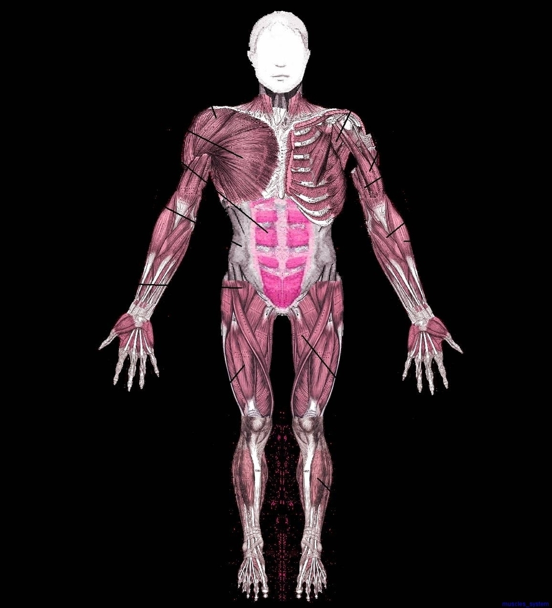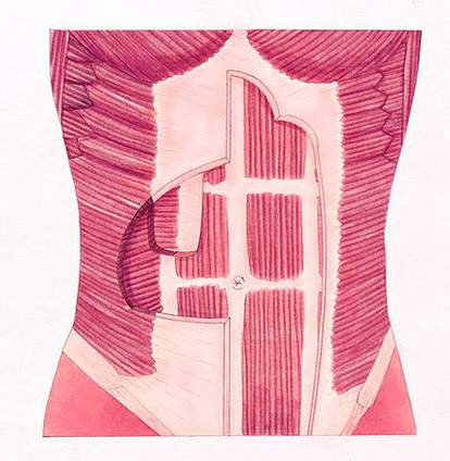


Anatomy Of The Muscles Image
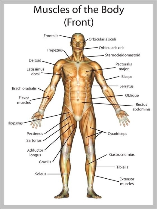
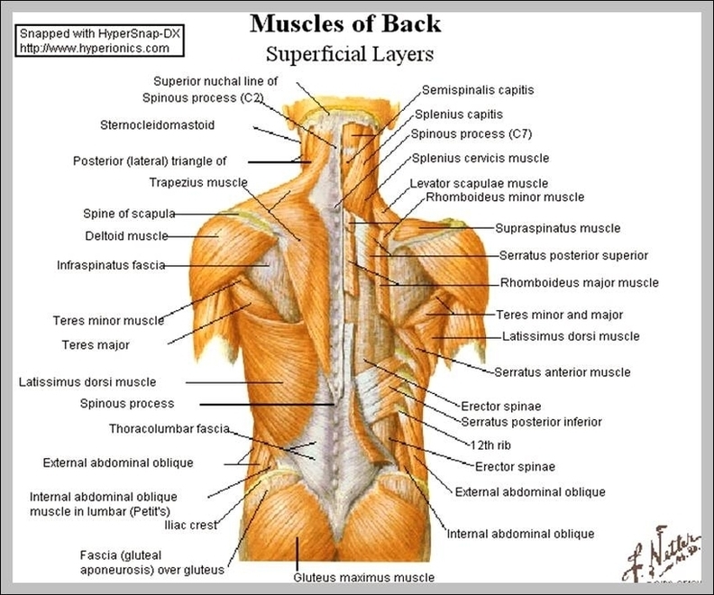
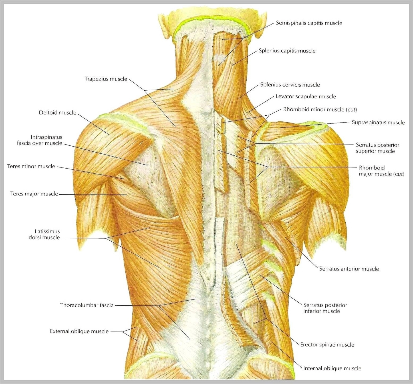
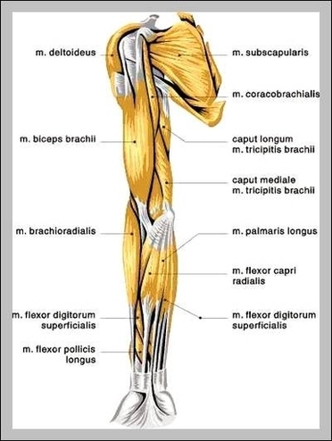
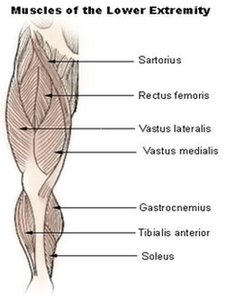
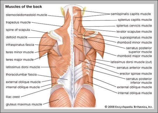
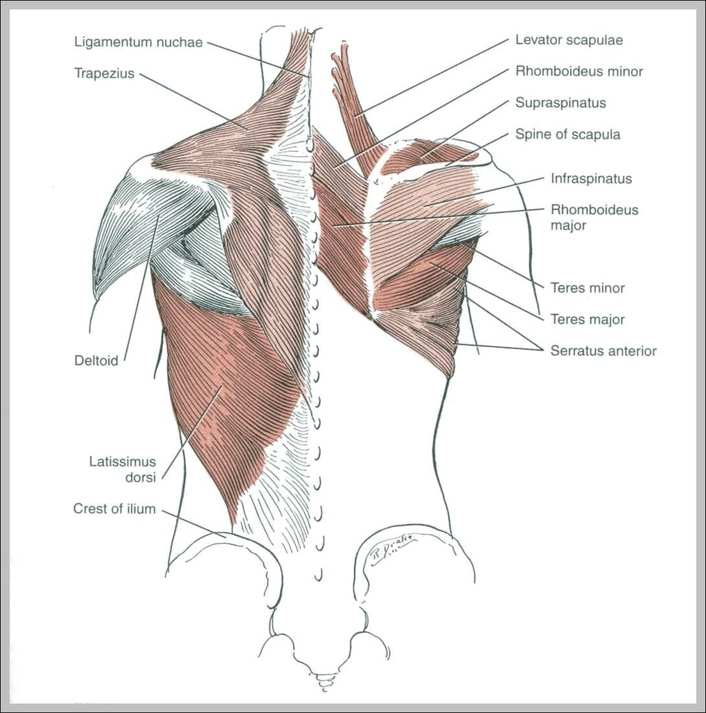
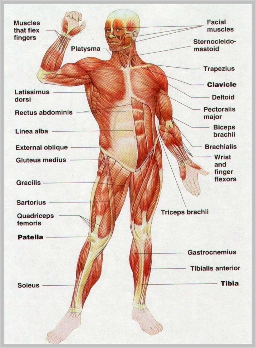
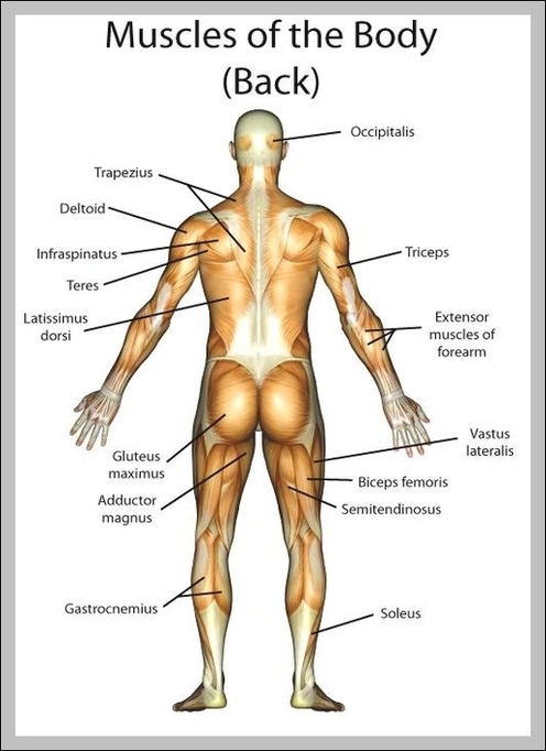
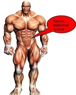
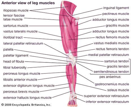
128,412 muscle anatomy stock photos, vectors, and illustrations are available royalty-free. Forearms – Anatomy Muscles Rhomboid minor and rhomboid major, levator scapulae and latissimus dorsi muscles – didactic board of anatomy of human bony and muscular system, posterior view Abs View Diagram Anatomy Of The Muscles Image