Diagram Of Anterior Leg Muscles Anterior View Madell Online Image

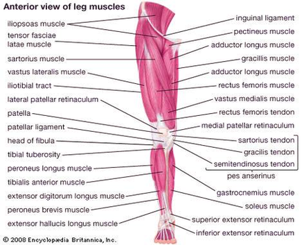
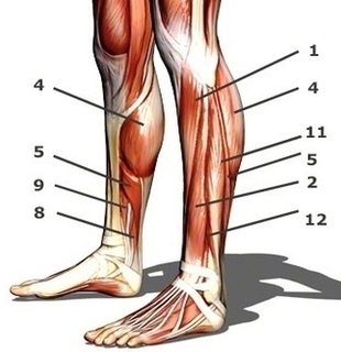
The anterior muscles of the leg. When studying the muscles of the leg, we can examine them by four primary groupings, which are the anterior, fibular/lateral, superficial posterior and deep posterior compartments. These are defined by intermuscular septa and surrounded by the deep fascia of the leg.

Tibialis anterior muscle This muscle is the most anterior and medial of all four anterior leg muscles. It originates from the proximal portion of the leg, precisely, from the lateral tibial condyle and proximal half of the tibial shaft, in addition to the adjacent portion of the interosseous membrane.

Do you visualise all of the muscles from the thigh down to the ankle? In fact, the muscles of the leg refer to the muscles found in the region between the knee and foot . In this article, we’re going to be teaching you about every last one of them, including their individual names, origins, insertions, innervations and functions.

Diagram Of Anterior Leg Muscles Anterior View Madell Online Image Diagram - Chart - diagrams and charts with labels. This diagram depicts Diagram Of Anterior Leg Muscles Anterior View Madell Online Image

Diagram Of Anterior Leg Muscles Anterior View Madell Online Image

Anatomy Of The Human Eye Cross Section View Image

Eye – Cross Section. A human eyeball cross section showing the following structures: iris, anterior limiting membrane, posterior limiting membrane, tendon of Eye anatomy, Human eye cross section physiology, Model of cornea and lens for ophthalmologist. Eye anatomy, Human eye cross section physiology, Model of cornea Enlarged anatomical eye model.

A human eyeball cross section showing the following structures: iris, anterior limiting membrane, posterior limiting membrane, tendon of Eye anatomy, Human eye cross section physiology, Model of cornea and lens for ophthalmologist.

Cross section of vitreous anatomy. A, Cross section diagram of the eye with emphasis on the anatomical features of the vitreous. The vitreous is most firmly attached to the retina at the vitreous base, and it also has adhesions at the optic nerve, along vessels, at the fovea, and to the posterior lens capsule.

Anatomy Of The Human Eye Cross Section View Image Diagram - Chart - diagrams and charts with labels. This diagram depicts Anatomy Of The Human Eye Cross Section View Image

Anatomy Of The Human Eye Cross Section View Image

Ankle Anterior View Radiograph Large Image

Radiographic analysis of the ankle often includes three views: the anteroposterior view, the internal oblique view, and the direct lateral view (15). The anteroposterior view helps assess the ankle mortise through the lateral portions of the talus and tibiotalar joint overlap with the lateral malleolus (16).

A P view Lateral view Internal oblique view Three views of the ankle are mandatory for the evaluation of the ankle following trauma. Some fractures of the medial malleolus may not be apparent on the AP and lateral views but are readily visible on the internal oblique projection.

The AP view (Figure 11-1 A) shows the ankle mortise; however, the lateral margin of the talus and lateral portions of the joint are obscured by the underlying lateral malleolus. Soft tissue swelling seen about the medial and lateral malleolus serves as a useful clue to the presence of an underlying acute fracture. FIGURE 11-1

Ankle Anterior View Radiograph Large Image Diagram - Chart - diagrams and charts with labels. This diagram depicts Ankle Anterior View Radiograph Large Image

Ankle Anterior View Radiograph Large Image

Bottom View Of Foot Bones Image

When one looks at the anatomy of the foot, they would realize that the foot has a complex mechanical and structural architecture. The ankle joint is the shock absorber of the foot. Apart from 28 bones, 33 joints, muscles, ligaments, and about 100 foot tendons make the foot. The diagram of bones in the ankle and foot is given below:

foot skeleton picture. Like already mentioned, the hindfoot is the posterior part of the foot. Its made up of 4 bones; The calcaneus, talus, fibula, and tibia bones. 1. The tibia bone. The tibia is one of the 2 bones that make up the leg. It extends from your knee joint upwards to the ankle joint downwards.

The forefoot is the furthermost part of the foot and consist of metatarsal bones and phalanges that make up your toes. The foot bones are generally grouped into tarsal, metatarsal and phalanges.

Bottom View Of Foot Bones Image Diagram - Chart - diagrams and charts with labels. This diagram depicts Bottom View Of Foot Bones Image

Bottom View Of Foot Bones Image

Wrist Radiography Ap View Imaios Anatomy En Medical Image

The 3D images of the wrist joint and carpal bones are three-dimensional reconstructions obtained from a scanner. 340 anatomical structures of the wrist were labeled, accessible on “Anatomical parts”: General anatomy: the different regions of the wrist and hand (carpal, metacarpal, hypothenar and thenar eminence).

Anatomy, medical imaging and e-learning for healthcare professionals – IMAIOS Anatomy atlas e-Anatomy and medical imaging learning resources available on the web and mobile (Android and iOS) ×Your email address is not verified.

Plain frontal and side-view X-Rays of the wrist show the lower extremities of the radius and ulna, the radiocarpal joint, the carpal bones (scaphoid, capitate, trapezium, trapezoid, hamate, lunate, pisiform and triquetral) and the carpometacarpal joints. Anatomy atlas , Radiographs , Wrist : Anterior-posterior view

Wrist Radiography Ap View Imaios Anatomy En Medical Image Diagram - Chart - diagrams and charts with labels. This diagram depicts Wrist Radiography Ap View Imaios Anatomy En Medical Image

Wrist Radiography Ap View Imaios Anatomy En Medical Image

Diagram Of Human Anatomy Mediastinum Left View Illustration Large Image

Nerves: details the nervous anatomy of the mediastinum including notably the route of the phrenic and vagus nerves as well as the anatomy of the sympathetic trunk at the thoracic level. Heart: the external morphology of the heart in the mediastinum and the various chambers of the heart on the anatomical sections are detailed on these diagrams.

The superior mediastinum contains three visceral organs including the esophagus, the trachea and remnants of the thymus. The nerves that run through this area are three and there are also two different nervous plexuses.

Middle Inferior Mediastinum. The middle inferior mediastinum contains a single nerve which is the phrenic nerve, the heart and the pericardium. The vessels present include: the ascending aorta. the pulmonary trunk. the superior vena cava. the pericardiacoophrenic artery.

Diagram Of Human Anatomy Mediastinum Left View Illustration Large Image Diagram - Chart - diagrams and charts with labels. This diagram depicts Diagram Of Human Anatomy Mediastinum Left View Illustration Large Image

Diagram Of Human Anatomy Mediastinum Left View Illustration Large Image

Calf Muscles Side View Image

2,195 female muscle calves stock photos and images available, or start a new search to explore more stock photos and images. Leg cramps during pregnancy. Closeup of hands massaging swollen foot while sitting on sofa female muscle calves stock pictures, royalty-free photos & images

The calf is defined largely as the three headed calf muscle (triceps surae muscle). In anatomy, we actually talk about the lower leg muscles and divide them into the following categories:

The calf muscle, on the back of the lower leg, is actually made up of two muscles: The gastrocnemius is the larger calf muscle, forming the bulge visible beneath the skin. The gastrocnemius has two parts or “heads,” which together create its diamond shape. The soleus is a smaller, flat muscle that lies underneath the gastrocnemius muscle.

Calf Muscles Side View Image Diagram - Chart - diagrams and charts with labels. This diagram depicts Calf Muscles Side View Image

Calf Muscles Side View Image