
c8dee1f3 f2c7 4b62 9102 abea4758ca25

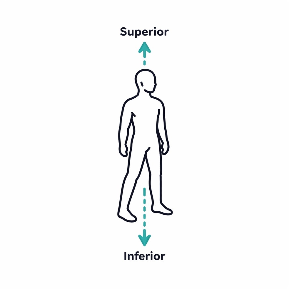
Anatomical planes, including sagittal, coronal, and transverse planes, are reference divisions of the human body used to describe location and orientation of structures. Knowledge of these planes is essential for clinicians, surgeons, radiologists, and medical students in imaging interpretation, surgical View Diagram The Anatomical Planes of the Human Body

The supratrochlear nerve, a branch of the ophthalmic division of the trigeminal nerve, supplies the skin of the medial forehead and upper eyelid. Knowledge of its course is essential for neurologists, surgeons, and medical students in performing nerve blocks, trauma View Diagram Course of the Supratrochlear Nerve
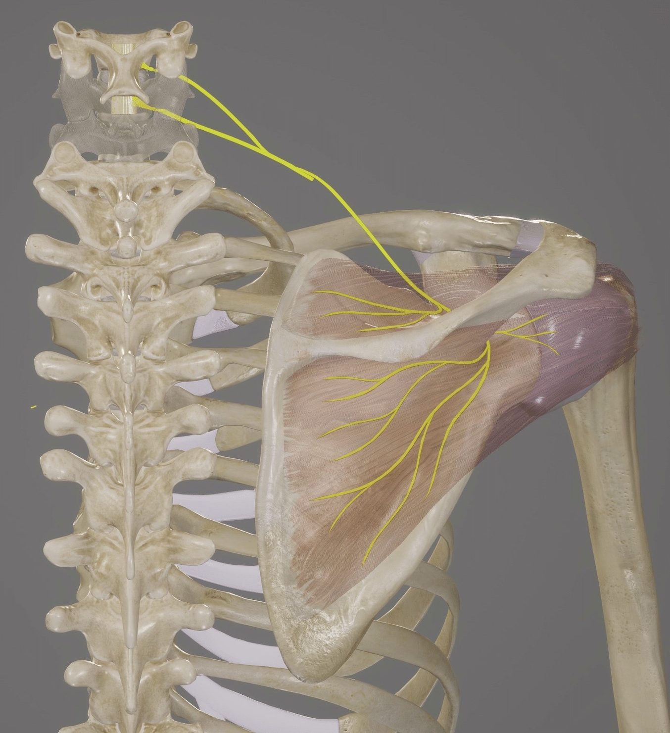
The suprascapular nerve arises from the upper trunk of the brachial plexus and supplies the supraspinatus and infraspinatus muscles. Knowledge of its course is essential for orthopedic surgeons, neurologists, and medical students in diagnosing entrapment syndromes, managing shoulder injuries, or View Diagram Suprascapular Nerve
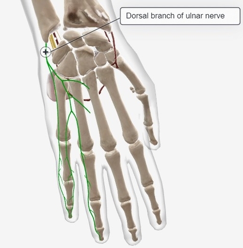
The dorsal branch of the ulnar nerve provides sensory innervation to the dorsoulnar aspect of the hand and motor supply to intrinsic hand muscles. Knowledge of its course is essential for surgeons, neurologists, and medical students in managing trauma, nerve View Diagram Dorsal Branch of the Ulnar Nerve
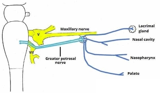
The pterygopalatine ganglion is a parasympathetic ganglion located in the pterygopalatine fossa. It receives preganglionic fibers from the facial nerve and sends postganglionic fibers to the lacrimal gland, nasal cavity, palate, and pharynx. The ganglion is a relay station for View Diagram The Pterygopalatine Ganglion and Branches
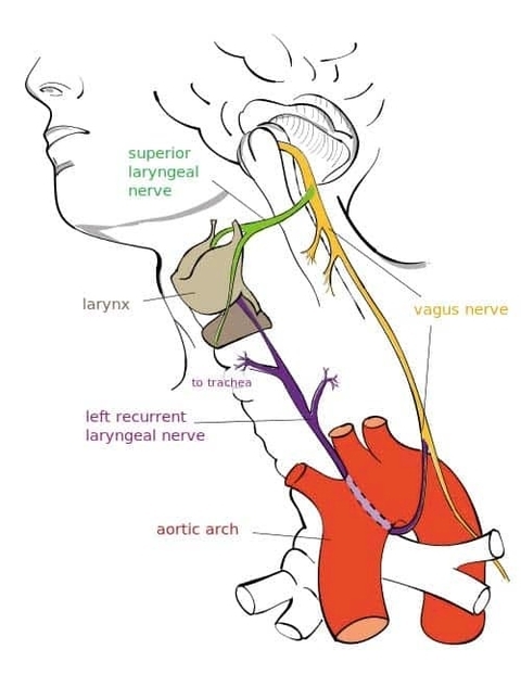
The vagus nerve is the tenth cranial nerve and has a wide distribution. It exits the skull through the jugular foramen and descends within the carotid sheath. In the thorax, it gives branches to the heart and lungs before contributing View Diagram Overview of the Major Branches and Anatomical Course of the Vagus Nerve
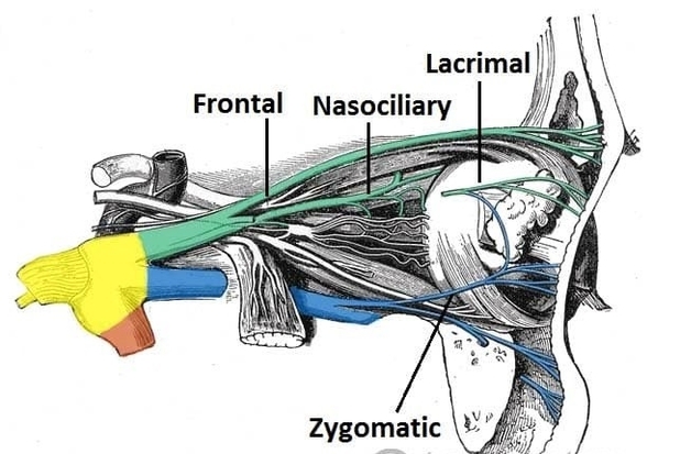
The ophthalmic nerve is the first division of the trigeminal nerve (cranial nerve V1) and provides sensory innervation to the upper face and orbit. This anatomy diagram of the anatomical course of the ophthalmic nerve illustrates its origin from the View Diagram Anatomical Course of the Ophthalmic Nerve
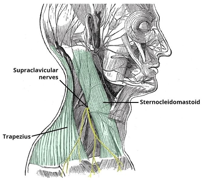
The supraclavicular nerves arise from the cervical plexus (C3C4) and supply the skin over the shoulder, clavicle, and upper chest. They are important in sensory innervation and commonly targeted in regional anesthesia. Knowledge of their course is essential in surgery, View Diagram Supraclavicular Nerves
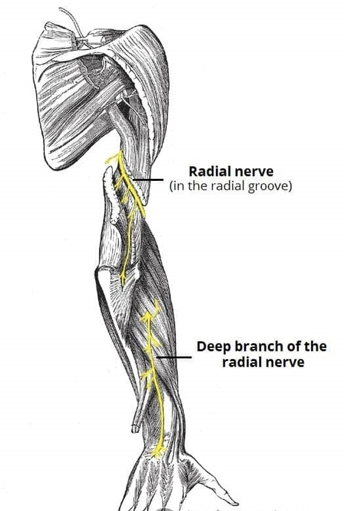
The radial nerve is a major peripheral nerve of the upper limb responsible for extension of the elbow, wrist, and fingers. This anatomy diagram of the anatomical course of the radial nerve traces its origin from the posterior cord of View Diagram Anatomical Course of the Radial Nerve
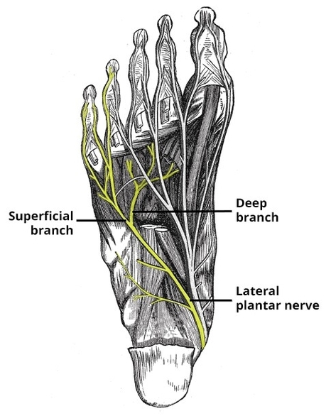
The lateral plantar nerve, a branch of the tibial nerve, courses along the lateral aspect of the sole, supplying intrinsic foot muscles and sensory innervation to the lateral toes. Knowledge of its course and branches is essential for orthopedic surgeons, View Diagram Lateral Plantar Nerve Course Branches

The auriculotemporal nerve follows a course from the mandibular nerve, supplying sensation to the temple, external ear, and temporomandibular joint. It also carries parasympathetic fibers to the parotid gland. Understanding its pathway is critical for dentists, surgeons, and medical students View Diagram Auriculotemporal Nerve Course Supply
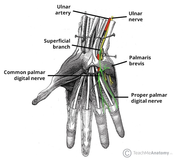
The superficial branch of the ulnar nerve divides into sensory and motor branches in the hand. Sensory branches supply the palmar and dorsal surfaces of the medial one and a half fingers, while motor fibers innervate intrinsic hand muscles, including View Diagram Superficial Branch of Ulnar Nerve Sensory and Motor Branches
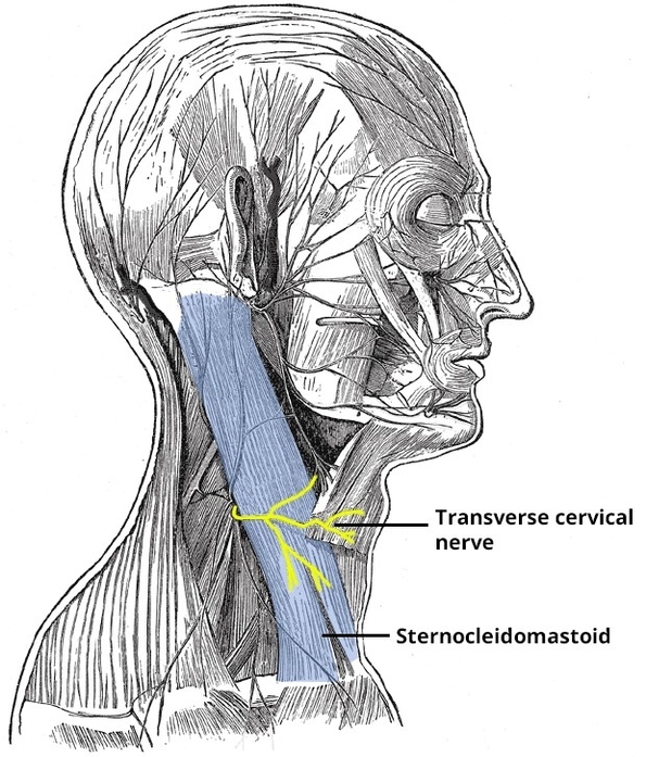
The transverse cervical nerve arises from the cervical plexus (C2C3). It emerges at the posterior border of the sternocleidomastoid and crosses the neck horizontally. It provides sensory innervation to the anterior neck. It is often encountered during surgical procedures in View Diagram Transverse Cervical Nerve
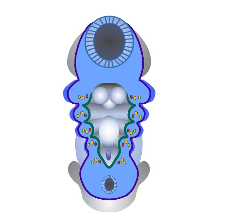
The pharyngeal arches are embryological structures that give rise to the face, neck, and pharyngeal components, including bones, muscles, and vessels. Each arch has a specific cranial nerve and skeletal contribution. Knowledge of pharyngeal arch development is essential for embryologists, View Diagram Pharyngeal Arches
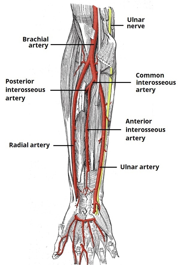
The radial and ulnar arteries are the main vessels supplying the forearm and hand. This anatomy diagram of the arterial supply to the forearm illustrates their origin from the brachial artery and course along the lateral and medial forearm. It View Diagram Arterial Supply to the Forearm Radial and Ulnar Arteries
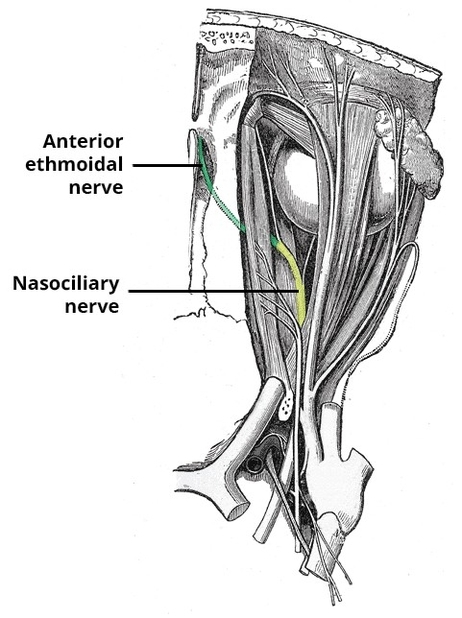
The anterior ethmoidal nerve is a branch of the nasociliary nerve that supplies the nasal cavity. This anatomy diagram of the anterior ethmoidal nerve shows its origin within the orbit, passage through the anterior ethmoidal foramen, and distribution to the View Diagram Anterior Ethmoidal Nerve

Pharyngeal nerves arise from the cranial nerves, primarily the glossopharyngeal and vagus nerves, and supply the pharyngeal muscles and mucosa. They are essential for swallowing, phonation, and sensory input from the pharynx. The course of these nerves passes through the View Diagram Pharyngeal Nerve Course Supply
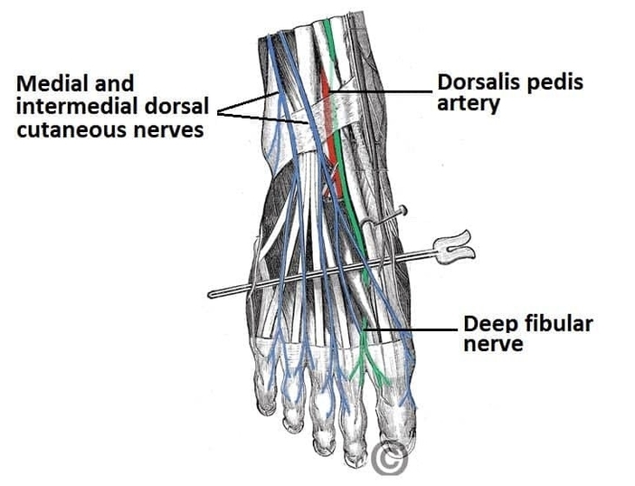
The nerves of the dorsum of the foot provide sensory innervation to the skin of the dorsal surface. This anatomy diagram of the anatomy of the nerves of the dorsum of the foot highlights the superficial and deep fibular nerves. View Diagram Anatomy of the Nerves of the Dorsum of the Foot Superficial and Deep Fibular Nerves
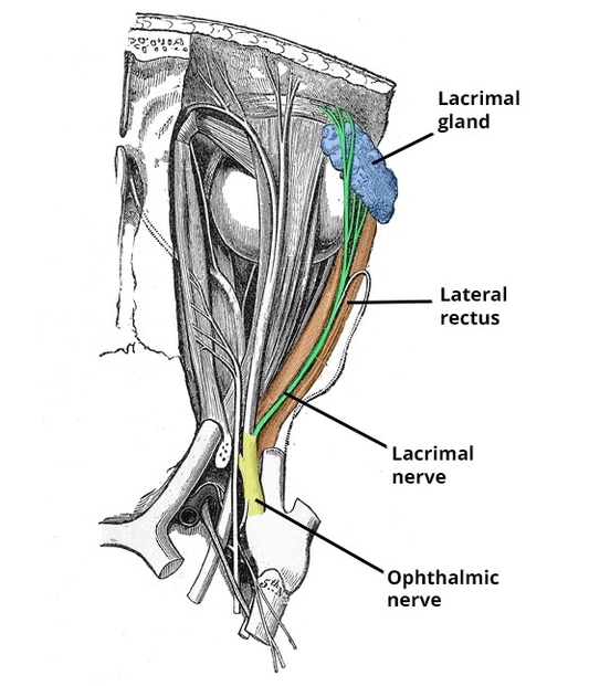
The lacrimal nerve, a branch of the ophthalmic division of the trigeminal nerve, provides sensory innervation to the lateral upper eyelid and conjunctiva. Knowledge of its course is essential for ophthalmologists, plastic surgeons, and medical students in eyelid surgery, trauma View Diagram Course of the Lacrimal Nerve