Kaposis Sacroma Lesions
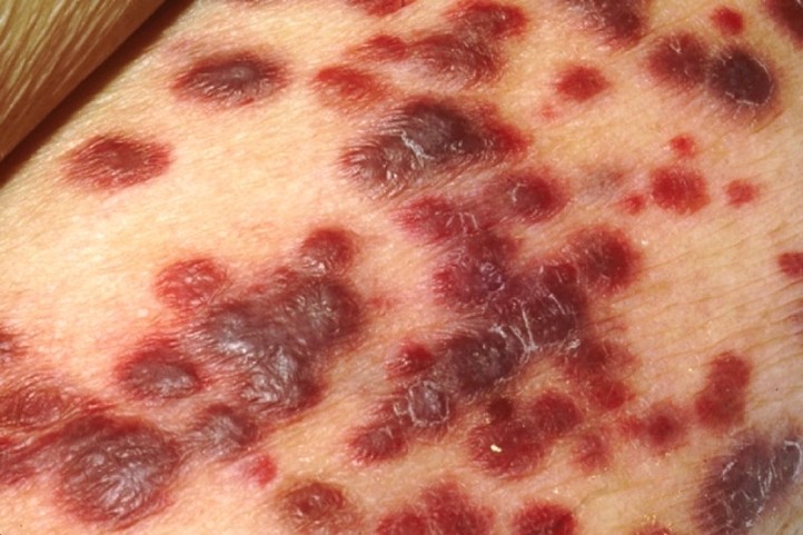
Kaposis Sarcoma Lesions: Kaposis sarcoma lesions are vascular tumors caused by human herpesvirus 8, often seen in immunocompromised individuals and appearing as purple or dark skin patches.