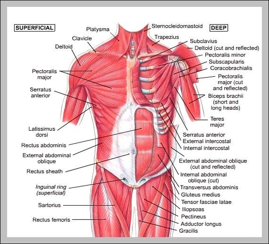
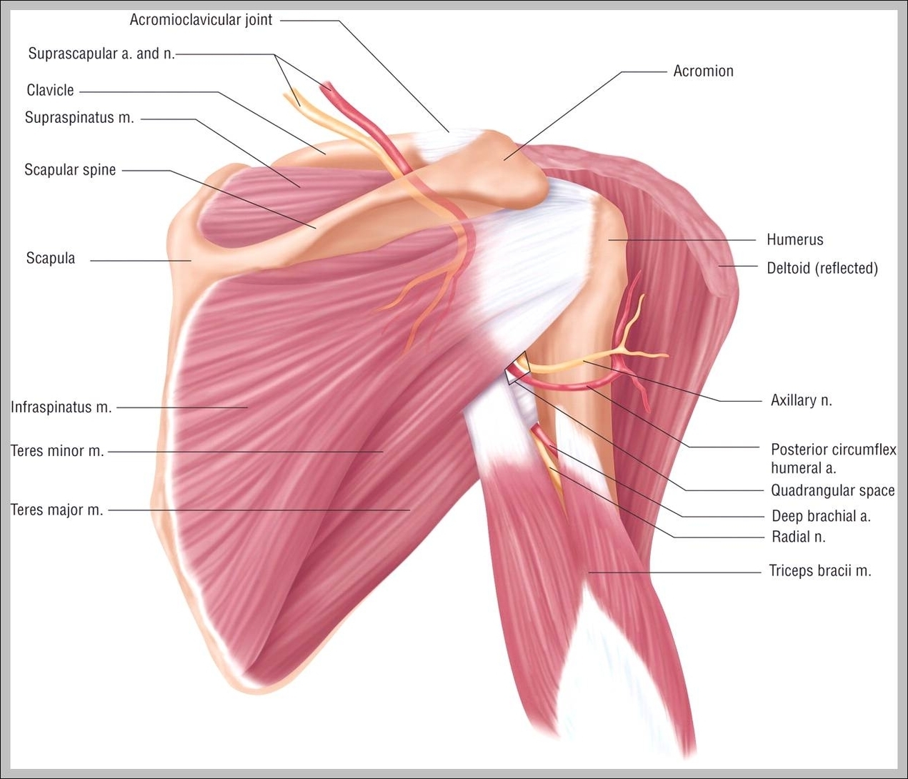
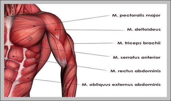
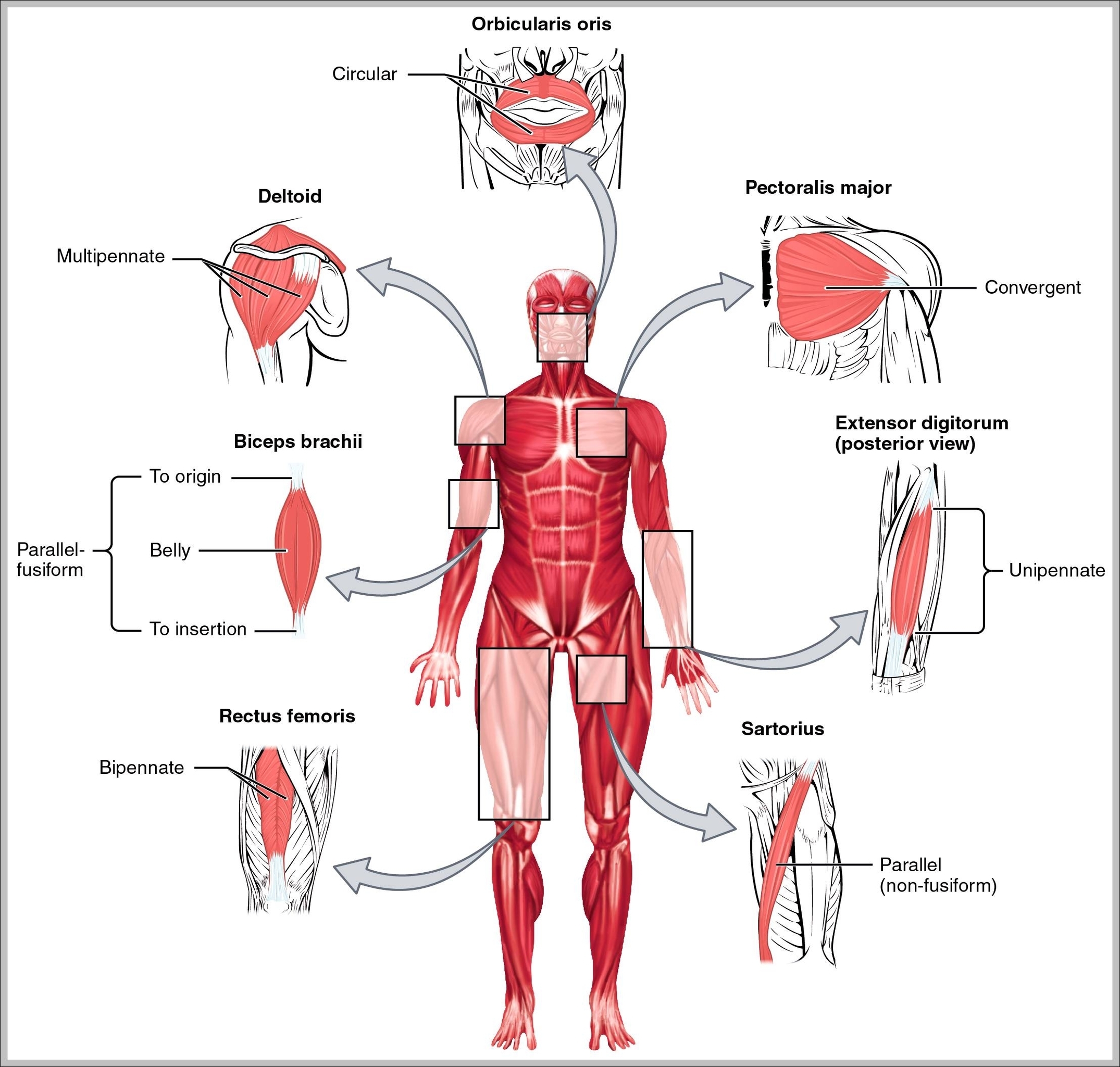
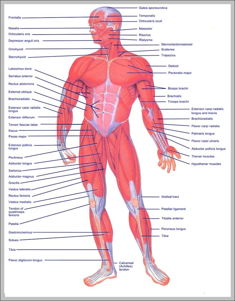
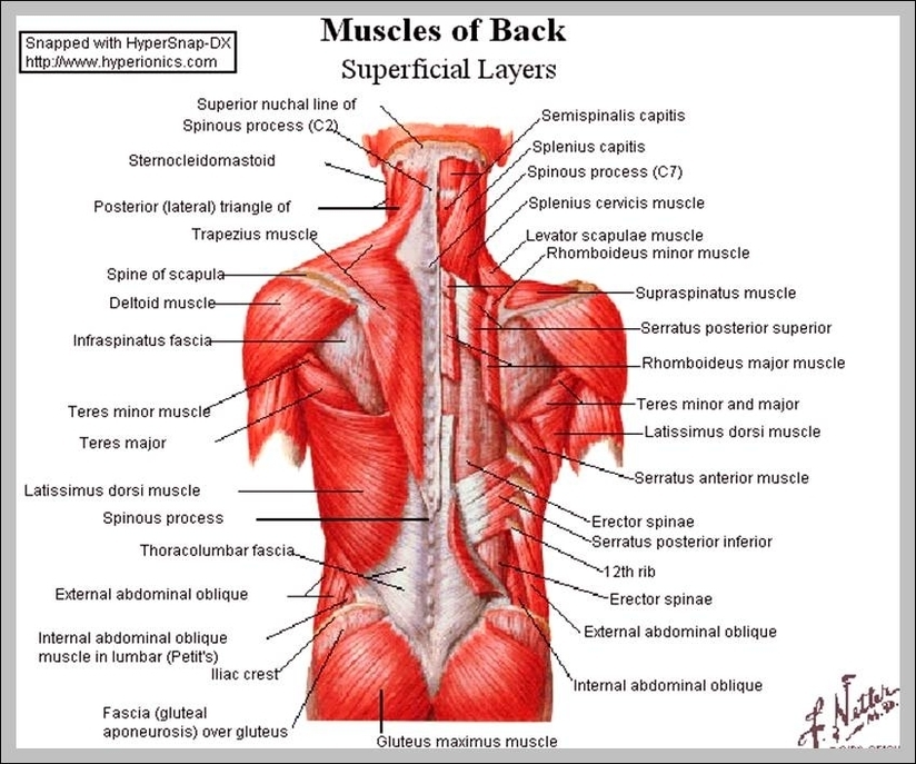
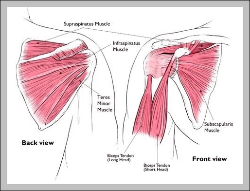
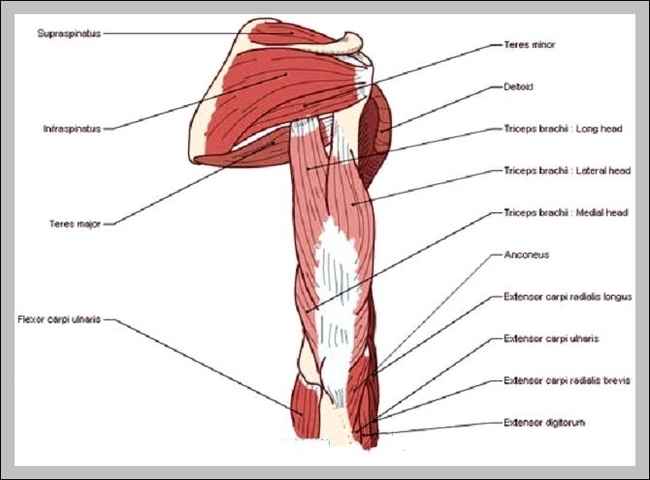
Skeletal Muscles Diagram
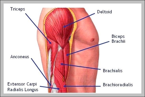
Attached to the bones of the skeletal system are about 700 named muscles that make up roughly half of a person’s body weight. Each of these muscles is a discrete organ constructed of skeletal muscle tissue, blood vessels, tendons, and View Diagram Skeletal Muscles Diagram