
Tag Archives: joint
Synovial Joint Diagram Image
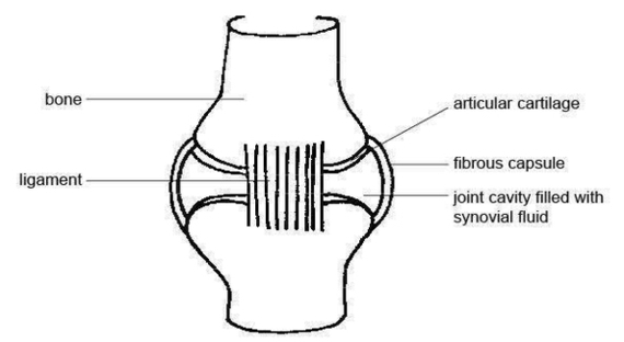
The structure of a synovial joint is demonstrated by a diagram in which the articulating bones are surrounded by the articular capsule, which comprises an exterior fibrous capsule and an interior synovial membrane. Start studying label the synovial joint. Learn View Diagram Synovial Joint Diagram Image
Hip Joint Muscles Image

hip joint anatomy images 19,596 hip joint anatomy stock photos, vectors, and illustrations are available royalty-free. See hip joint anatomy stock video clips Muscles of the Hip. The hip joint is one of the most flexible joints in the entire View Diagram Hip Joint Muscles Image
Diagram Illu Synovial Joint Image
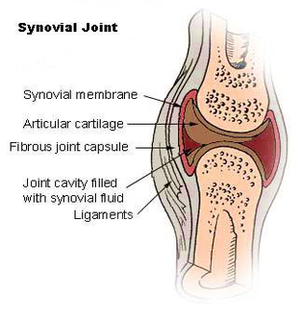
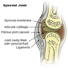
The structure of a synovial joint is demonstrated by a diagram in which the articulating bones are surrounded by the articular capsule, which comprises an exterior fibrous capsule and an interior synovial membrane. Start studying label the synovial joint. Learn View Diagram Diagram Illu Synovial Joint Image
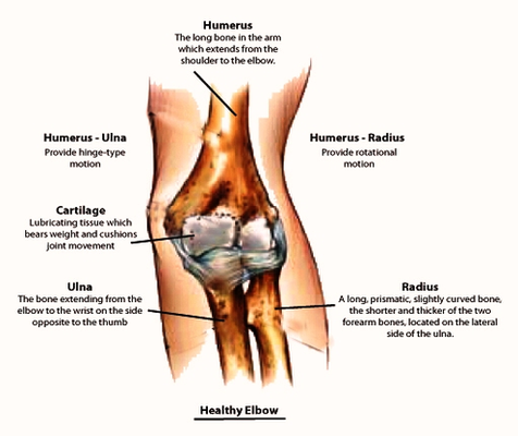
Elbow Joint Anatomy Image
Ankle Joint Anatomy On Healthfavo Image
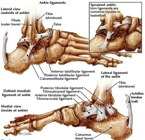
The ankle joint is an important joint in the human body, having a wide range of movements and consisting of different bones and ligaments. Learn now! This is an article covering the anatomy of the Tibia – interaction with the View Diagram Ankle Joint Anatomy On Healthfavo Image
Elbow Joint Diagram Image
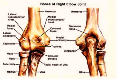
The students can follow these steps to make their elbow joint diagram: Step 1: The student should first draw the upper part of the front part of the elbow that has a pulley-like structure, trochlea. After that, they should create View Diagram Elbow Joint Diagram Image
Anatomy Glenohumeral Joint Shoulder Ligaments En Medical Image
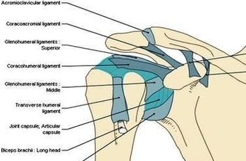
Glenohumeral ligaments (superior, middle and inferior) – the joint capsule is formed by this group of ligaments connecting the humerus to the glenoid fossa. They are the main source of stability for the shoulder, holding it in place and preventing View Diagram Anatomy Glenohumeral Joint Shoulder Ligaments En Medical Image
Muscles Of The Shoulder Joint
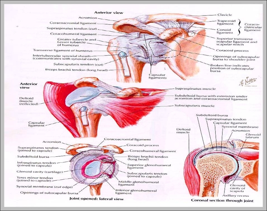
Muscles Of The Shoulder Joint: The muscles around the shoulder joint include the deltoid, rotator cuff, trapezius, and pectoralis, responsible for a wide range of arm and shoulder movements.
Hip Joint Type

Hip Joint Type: The hip is a ball-and-socket joint, allowing multidirectional movement and rotation, formed by the femoral head and the acetabulum of the pelvis.
Hinge Joint Movement
Hinge Joint Movement: Hinge joints, such as the elbow and knee, allow movement in one directionflexion and extensionsimilar to the opening and closing of a door.