Connective Tissue1 Diagram1 Image

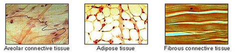
20,642 connective tissue stock photos and images available, or search for connective tissue disease or connective tissue massage to find more great stock photos and pictures. Areolar connective tissue under the microscope view.

The primary elements of connective tissue include a ground substance, fibers, and cells. Loose connective tissue holds organs in place and attaches epithelial tissue to other underlying tissues.

Specialised Connective Tissues. Type # 1. Loose Connective Tissue: This tissue is most widely distributed connective tissue in the animal body. It is named so because it takes the form of fine threads crossing each other in every direction leaving small spaces called areolae.

Connective Tissue1 Diagram1 Image Diagram - Chart - diagrams and charts with labels. This diagram depicts Connective Tissue1 Diagram1 Image

Connective Tissue1 Diagram1 Image

Crown And Bridge Image1 Image

They have two crowns – one on either end – and a bridge of replacement teeth that rest in the area of the gums where there is tooth loss. The crowns at the ends of a bridge can fuse to existing teeth (that need to be filed down by a dental professional to fit correctly), or your dental professional can attach them to dental implants.

For a bridge, your dental professional will place caps on filed-down teeth or dental implants at the outer edges of your area of loss. Replacement teeth (called pontics) span the space between the caps where the teeth are missing. Just like with dental crowns, you have a choice of materials available to you for your bridge.

A dental crown can cap an individual’s damaged or decaying tooth or be placed on top of a dental implant to replace a missing tooth. Dental implants are metal fixtures, surgically fastened to your jawbone below the gums.

Crown And Bridge Image1 Image Diagram - Chart - diagrams and charts with labels. This diagram depicts Crown And Bridge Image1 Image

Crown And Bridge Image1 Image

Cyclacel Cell Cycle Diagram1 Image

The phases are: 1. G1 (gap1) phase 2. S (synthesis) phase 3. G2 (gap 2) phase 4. M (mitosis) phase. Cell Cycle: Phase # 1.

Our strategy is to build a diversified portfolio of novel drug candidates to address areas of high unmet medical needs. Cyclacel is a pioneer in the field of cell cycle biology. Our vision is to improve patient healthcare by translating cancer biology into medicines.

The G 1 phase is set in immediately after the cell division. It is characterised by a change in the chromosome from the condensed mitotic state to the more extended interphase state and by a series of metabolic events leading to initiation of DNA replication. G, phase of cell cycle varies in length from cell to cell within the same cell population.

Cyclacel Cell Cycle Diagram1 Image Diagram - Chart - diagrams and charts with labels. This diagram depicts Cyclacel Cell Cycle Diagram1 Image

Cyclacel Cell Cycle Diagram1 Image

Dep The Abo Blood Group Diagram1 Image

It is not to be confused with Typo or Type 0. The ABO blood group system is used to denote the presence of one, both, or neither of the A and B antigens on erythrocytes. In human blood transfusions it is the most important of the 38 different blood type (or group) classification systems currently recognized.

ABO antibodies in the serum are formed naturally. when the immune system encounters the “missing” ABO blood group antigens in foods or in micro-organisms. This happens at an early age because sugars that are identical to, or very similar to, the ABO blood group antigens are found

The four basic ABO phenotypes are O, A, B, and AB. After it was found that blood group A RBCs reacted differently to a particular antibody (later called anti-A1), the blood group was divided into two phenotypes, A 1 and A 2.

Dep The Abo Blood Group Diagram1 Image Diagram - Chart - diagrams and charts with labels. This diagram depicts Dep The Abo Blood Group Diagram1 Image

Dep The Abo Blood Group Diagram1 Image

Ankle Anatomy1 Image

1,365 ankle anatomy stock photos and images available, or search for foot and ankle anatomy to find more great stock photos and pictures.

The anatomy of the foot is incredibly complex. This introduction to the anatomy of the foot and ankle will be very general and highlight the most relevant structures. The ankle joint or tibiotalar joint is formed where the top of the talus (the uppermost bone in the foot) and the tibia (shin bone) and fibula meet.

There are many different muscles and ligaments in the ankle, which gives the ankle its strength, flexibility, and range of motion. Save. Anterior and posterior ankle ligaments. Ligaments are a type of soft tissue that is made up mostly of collagen.

Ankle Anatomy1 Image Diagram - Chart - diagrams and charts with labels. This diagram depicts Ankle Anatomy1 Image

Ankle Anatomy1 Image

Upper Extremity Diagram1 Image

The forearm is the portion between the elbow and wrist. The thigh is the portion of the lower extremity between the hip and knee, and the calf is the portion between the knee and ankle. The normal arterial anatomy of the upper extremity is depicted graphically in Figure 13-1.

The next chapter is about the osteology of the upper limb, with all its anatomical structures, muscle insertions and ligaments of the bones: scapula, clavicle, humerus, ulna, radius, carpal bones, metacarpals and phalanges of the fingers.

The final chapter presents anatomical charts of anatomical sections of the upper limb: the axilla and the deltoid region in axial and coronal and axial sections of the arm, the elbow, forearm, wrist, carpal and metacarpal regions.

Upper Extremity Diagram1 Image Diagram - Chart - diagrams and charts with labels. This diagram depicts Upper Extremity Diagram1 Image

Upper Extremity Diagram1 Image

Brain Diagram1 Image

26,763 brain diagram stock photos, vectors, and illustrations are available royalty-free.

WebMD’s Brain Anatomy Page provides a detailed diagram and definition of the brain including its function, parts, and conditions that affect it. Skip to main content Check Your Symptoms Find A Doctor Find A Dentist Connect to Care Find Lowest Drug Prices Health A-Z Health A-Z Common Conditions ADD/ADHD Allergies

Labeled Diagrams of the Human Brain. Central Core. The central core consists of the thalamus, pons, cerebellum, reticular formation and medulla. These five regions are the central areas that regulate breathing, pulse, arousal, balance, sleep and early stages of processing sensory information.

Brain Diagram1 Image Diagram - Chart - diagrams and charts with labels. This diagram depicts Brain Diagram1 Image

Brain Diagram1 Image scaled

Arm Anatomy1 Image

4,995 human arm anatomy stock photos and images available, or start a new search to explore more stock photos and images.

The human arm is divided into two main regions, the portion from the elbow to the wrist known as the forearm, and the segment from the shoulder to the elbow referred to as the arm. Arm muscle anatomy enables the arm to perform a variety of movements, including flexion, extension, pronation, and supination.

The muscles of the arms attach to the shoulder blade, upper arm bone ( humerus ), forearm bones ( radius and ulna ), wrist, fingers, and thumbs. These muscles control movement at the elbow, forearm, wrist, and fingers. When affected by injury or neuromuscular disorders, everyday tasks that require hand and arm use can be challenging.

Arm Anatomy1 Image Diagram - Chart - diagrams and charts with labels. This diagram depicts Arm Anatomy1 Image

Arm Anatomy1 Image

Human Body Diagram1 Image

43,556 human body diagram stock photos, vectors, and illustrations are available royalty-free.

internal organs and circulatory system Vector isolated illustration of human internal organs and circulatory system in man body. Stomach, liver, bladder, lung, kidney, heart, icon. Medical poster organs of the human body diagram stock illustrations Vector isolated illustration of human internal organs and circulatory system in man body.

A vast array of aspects concerning the human body have been comprehended; however, there are facets that await a treatment for thorough analysis. The head, neck, torso, a pair of arms and legs, respectively constitute the external view of the body, often described as the superficial, first-layer of the human body.

Human Body Diagram1 Image Diagram - Chart - diagrams and charts with labels. This diagram depicts Human Body Diagram1 Image

Human Body Diagram1 Image

Ear Anatomy1 Image

1,061 ear anatomy stock photos and images available, or search for anatomy model or muscle anatomy to find more great stock photos and pictures.

Inner Ear Anatomy The inner ear is where the sound waves are translated into types of electrical nerve impulses. Most of the hearing and balance content is located within the bony labyrinth. After the tympanic membrane, these are the nerves that most likely contribute to hearing impairment and may require treatment or medical services.

The ear has external, middle, and inner portions. The outer ear is called the pinna and is made of ridged cartilage covered by skin. Sound funnels through the pinna into the external auditory canal, a short tube that ends at the eardrum (tympanic membrane).

Ear Anatomy1 Image Diagram - Chart - diagrams and charts with labels. This diagram depicts Ear Anatomy1 Image

Ear Anatomy1 Image

Digestive System Diagram1 Image

9,435 diagram digestive system stock photos, vectors, and illustrations are available royalty-free.

In this system, the process of digestion has The human digestive system consists of the gastrointestinal tract plus the accessory organs of digestion.

English: The gastrointestinal tract, also called the digestive tract, alimentary canal, or gut, is the system of organs within multicellular animals that takes in food, digests it to extract energy and nutrients, and expels the remaining waste. 日本語: ヒトの 消化器 の図。

Digestive System Diagram1 Image Diagram - Chart - diagrams and charts with labels. This diagram depicts Digestive System Diagram1 Image

Digestive System Diagram1 Image

Muscles Diagram1 Image

128,412 muscle anatomy stock photos, vectors, and illustrations are available royalty-free.

Tibialis Anterior Muscle Tibialis Posterior Muscle Vastus Lateralis Muscle Vastus Medialis Muscle GENERAL Cross-Sections of Muscles of the Arm and Leg Muscle Cell Types Pronation/Supination Push/Pull Muscles Change Current View Angle Muscular System (Male View) Muscular System (Male Posterior View) Muscular System (Posterior View)

Finally, the direction in which the muscle fibers run can be used to identify a muscle. In the abdominal region, there are several sets of wide, flat muscles.

Muscles Diagram1 Image Diagram - Chart - diagrams and charts with labels. This diagram depicts Muscles Diagram1 Image

Muscles Diagram1 Image