Diagram Of Anterior Leg Muscles Anterior View Madell Online Image

The anterior muscles of the leg. When studying the muscles of the leg, we can examine them by four primary groupings, which are the anterior, fibular/lateral, superficial posterior and deep posterior compartments. These are defined by intermuscular septa and surrounded by the deep fascia of the leg.

Tibialis anterior muscle This muscle is the most anterior and medial of all four anterior leg muscles. It originates from the proximal portion of the leg, precisely, from the lateral tibial condyle and proximal half of the tibial shaft, in addition to the adjacent portion of the interosseous membrane.

Do you visualise all of the muscles from the thigh down to the ankle? In fact, the muscles of the leg refer to the muscles found in the region between the knee and foot . In this article, we’re going to be teaching you about every last one of them, including their individual names, origins, insertions, innervations and functions.

Diagram Of Anterior Leg Muscles Anterior View Madell Online Image Diagram - Chart - diagrams and charts with labels. This diagram depicts Diagram Of Anterior Leg Muscles Anterior View Madell Online Image

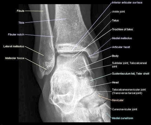
Diagram Of Anterior Leg Muscles Anterior View Madell Online Image

Ankle Anterior View Radiograph Large Image

Radiographic analysis of the ankle often includes three views: the anteroposterior view, the internal oblique view, and the direct lateral view (15). The anteroposterior view helps assess the ankle mortise through the lateral portions of the talus and tibiotalar joint overlap with the lateral malleolus (16).

A P view Lateral view Internal oblique view Three views of the ankle are mandatory for the evaluation of the ankle following trauma. Some fractures of the medial malleolus may not be apparent on the AP and lateral views but are readily visible on the internal oblique projection.

The AP view (Figure 11-1 A) shows the ankle mortise; however, the lateral margin of the talus and lateral portions of the joint are obscured by the underlying lateral malleolus. Soft tissue swelling seen about the medial and lateral malleolus serves as a useful clue to the presence of an underlying acute fracture. FIGURE 11-1

Ankle Anterior View Radiograph Large Image Diagram - Chart - diagrams and charts with labels. This diagram depicts Ankle Anterior View Radiograph Large Image

Ankle Anterior View Radiograph Large Image