Adam Ear Anatomy Image

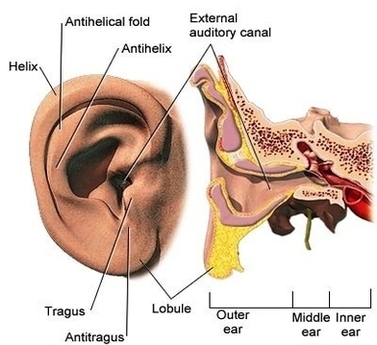
Adamimages.com is one of the world’s largest libraries of medical illustrations with nearly 30,000 detailed and medically accurate images ready for immediate download. our comprehensive image catalog covering disease states, injuries, conditions, tests, treatments, surgery procedures and human anatomy.

diagram of the anatomy of the human ear. Three ossicles: malleus, incus, and stapes (hammer, anvil, and stirrup). The ossicles directly couple sound energy from the ear drum to the oval window of the cochlea. Detailed illustration for educational, medical, biological, and scientific use.

The most comprehensive digital database of detailed anatomical images in the world, with the ability to identify over 20,000 anatomical structures from different body orientations (anterior, lateral, medial, posterior, lateral arm, medial arm).

Adam Ear Anatomy Image Diagram - Chart - diagrams and charts with labels. This diagram depicts Adam Ear Anatomy Image

Adam Ear Anatomy Image

Ankle Sprains Adam Image

A sprained ankle occurs when the ligaments are forced beyond their normal range of motion. Most sprained ankles involve injuries to the ligaments on the outer side of the ankle. Treatment for a sprained ankle depends on the severity of the injury.

Sprained ankle. Diagnosis. During a physical, your doctor will examine your ankle, foot and lower leg. The doctor will touch the skin around the injury to check for points of tenderness and move your foot to check the range of motion and to understand what positions cause discomfort or pain.

In the case of a severe sprain, a cast or walking boot may be necessary to immobilize the ankle while it heals. Once the swelling and pain is lessened enough to resume movement, your doctor will ask you to begin a series of exercises to restore your ankle’s range of motion, strength, flexibility and stability.

Ankle Sprains Adam Image Diagram - Chart - diagrams and charts with labels. This diagram depicts Ankle Sprains Adam Image

Ankle Sprains Adam Image