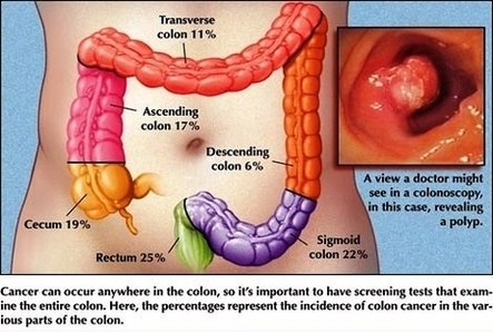
Colorectal Cancer Lg Image
4,035 colorectal cancer stock photos and images available, or search for colon cancer screening or colonoscopy to find more great stock photos and pictures. Doctor goes over a patient”s x-ray, screening for colon cancer. There is no single cause of View Diagram Colorectal Cancer Lg Image