Anatomy And Physiology Of Animals The Ear Image

This human anatomy diagram with labels depicts and explains the details and or parts of the Anatomy And Physiology Of Animals The Ear Image. Human anatomy diagrams and charts show internal organs, body systems, cells, conditions, sickness and symptoms information and/or tips to ensure one lives in good health.

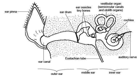
Anatomy and physiology of the canine ear The canine ear consists of the pinna, external ear canal, middle ear and inner ear. The external ear is composed of auricular and annular cartilage. The auricular cartilage of the pinna becomes funnel shaped at the opening of the external ear canal. The vertical ear canal runs for about 1 inch, then …

The ear canal contains protective hairs and ear wax ( cerumen ). The middle ear (tympanic cavity) lies behind the eardrum. The middle ear contains three small bones (ossicles) that transmit sound waves from the eardrum to the inner ear. The three bones are called the malleus, the incus, and the stapes.

Other than hearing, the main function is to maintain the balance of the body. The hair cells present in the inner ear of mammals help in sensing the position of the body, in accordance with gravity and maintain the equilibrium. Structure of ear comprises three main sections: the outer ear, middle ear and inner ear.

Anatomy And Physiology Of Animals The Ear Image

Anatomy And Physiology Of Animals The Ear Image


Tags: , ,