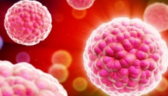
(Learn more about how cancer spreads .) Appearance —Under a microscope, normal cells and cancer cells may look quite different. In contrast to normal cells, cancer cells often exhibit much more variability in cell size—some are larger than normal and some are smaller than normal.
Cancer cells Concept of Education anatomy and Human lung tissue under microscope, The lungs is organs of the respiratory system in humans. Normal human cell reborn to cancer cells, and growing to malignant tumor. Human anatomy Prostate cancer cells, 3D illustration. Prostate cancer awareness image Human lung tissue under microscope view.
In addition, cancer cells often have an abnormal shape, both of the cell, and of the nucleus (the “brain” of the cell.) The nucleus appears both larger and darker than normal cells.
Cancer Cells Ve Image Diagram - Chart - diagrams and charts with labels. This diagram depicts Cancer Cells Ve Image

