Cub Cells Lesson Figure Image

This human anatomy diagram with labels depicts and explains the details and or parts of the Cub Cells Lesson Figure Image. Human anatomy diagrams and charts show internal organs, body systems, cells, conditions, sickness and symptoms information and/or tips to ensure one lives in good health.

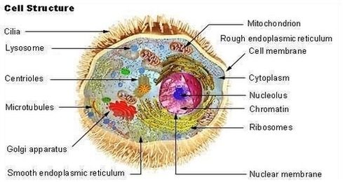
In the second lesson, students learn about the basics of cellular respiration. They also learn about the application of cellular respiration to engineering and bioremediation. The third lesson continues students’ education on cells in the human body and how (and why) engineers are involved in the research of stem cell behavior.

Worksheet/Drawing: Have the students draw and label their blood cell on the Blood Cell Model Worksheet; review their answers to gauge their mastery of the subject. Group Questions: During the activity, ask the groups discussion questions as directed in procedures above.

Make copies of Blood Cell Model Worksheet and Blood Cells Under a Microscope Worksheet (for optional section). Each student should have their own worksheet (s). Have all materials set out. If available, set up the microscopes with the blood cell slides. Be sure to focus the microscope for the clearest view.

Cub Cells Lesson Figure Image

Cub Cells Lesson Figure Image


Tags: , , ,