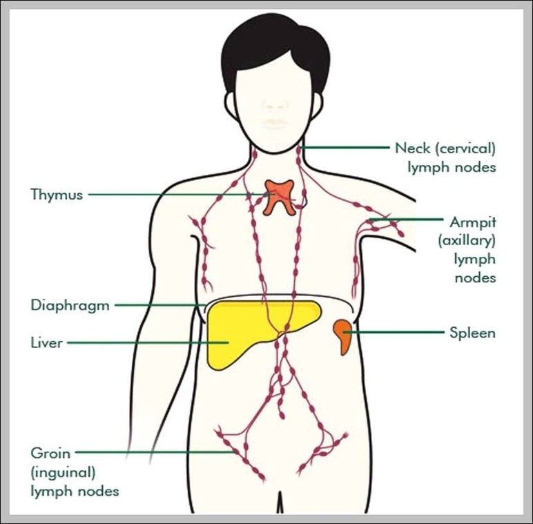
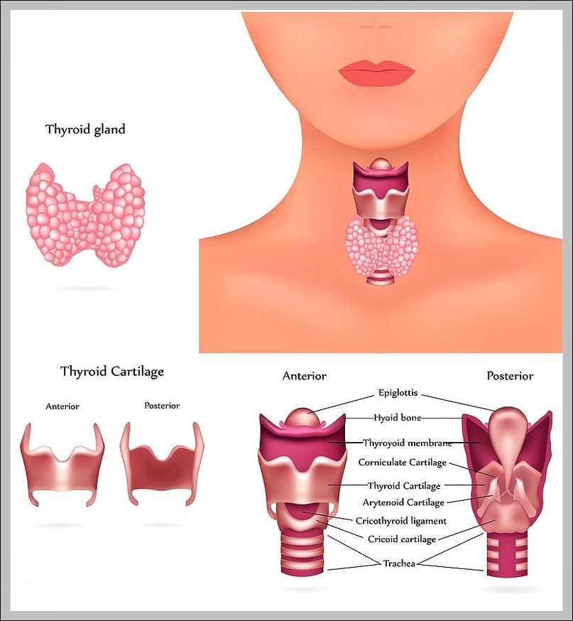
Thyroid Gland Scan Image
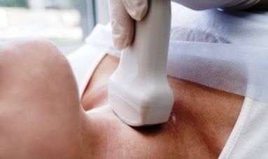
An ultrasound is a painless procedure that uses sound waves to generate images of the inside of your body. Your doctor will often use an ultrasound to create images of a fetus during pregnancy. A thyroid ultrasound is used to View Diagram Thyroid Gland Scan Image