Diagram Of Cancer Cells Dead Blood Dead Blood Image
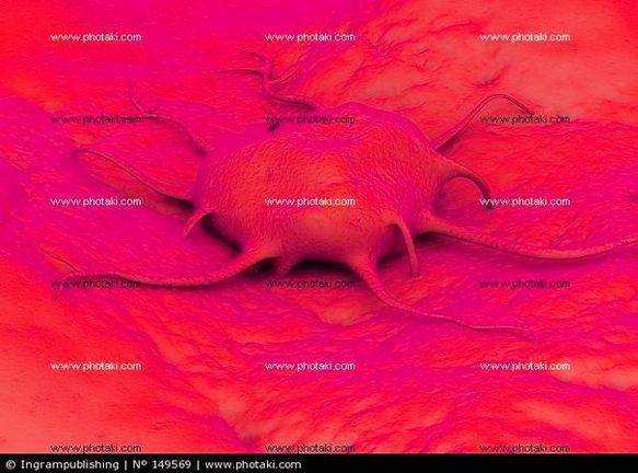
This human anatomy diagram with labels depicts and explains the details and or parts of the Diagram Of Cancer Cells Dead Blood Dead Blood Image. Human anatomy diagrams and charts show internal organs, body systems, cells, conditions, sickness and symptoms information and/or tips to ensure one lives in good health.

Dead cancer cells are as tasty to a phagocyte as any other type of dead cell. The dead cells cannot “re-enter the bloodstream” once they’ve been digested by macrophages, because the digestion process breaks down the chunks into individual chemical components (amino acids, lipids, sugars, minerals).

Blood Cancer Under the Microscope 1 Chronic Myeloid Leukemia. On the associated image of a peripheral blood smear of a patient,… 2 Hodgkin Lymphoma. This slide shows a case of Hodgkin lymphoma, sometimes called Hodgkin disease. 3 Human Immune Cell Killing a Cancer Cell. Here we see a killer T cell…

In this case, the scavengers are white blood cells whose job is to eat (literally) and digest dead things such as dead cells and cell debris. There are two main types of cells that do that: neutrophils and macrophages, both of which are called “phagocytes”. The neutrophils almost always come from the blood, and they get to the dead cells first.

Diagram Of Cancer Cells Dead Blood Dead Blood Image

Diagram Of Cancer Cells Dead Blood Dead Blood Image


Tags: , ,