Tag Archives: physiology
Anatomy And Physiology Of Animals The Ear Image
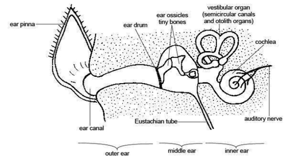
Anatomy and physiology of the canine ear The canine ear consists of the pinna, external ear canal, middle ear and inner ear. The external ear is composed of auricular and annular cartilage. The auricular cartilage of the pinna becomes funnel View Diagram Anatomy And Physiology Of Animals The Ear Image
Anatomy And Physiology Pictures Image
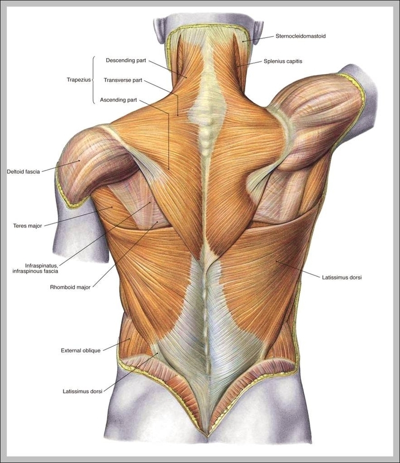
87,967 professional anatomy stock photos available royalty-free. Old vintage anatomy charts of the human body. Showing the skeletal system and various muscles, four figures in a row in different orientations SPINE Pain – Male Hurt Backbone isolated on white – View Diagram Anatomy And Physiology Pictures Image
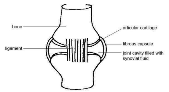
Anatomy And Physiology Of Animals Structure Of Muscle Image
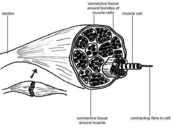
Here is a nice 50-questions quiz on animal muscles both as gross anatomy and in a microscope. This quiz; however, will focus on the gross anatomy of muscles. Muscle is a soft tissue found in most animals. They are primarly View Diagram Anatomy And Physiology Of Animals Structure Of Muscle Image
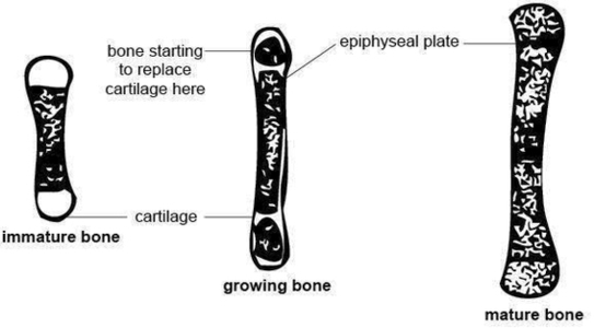
Anatomy And Physiology Of Animals Growing Bone1 Image

138Anatomy and physiology of domestic animals associated with bone formation, it can occur in other tissues. There are two general classes of bone formation. Intramembranous ossifi cation occurs when bone develops from a fi brous membrane. The fl at bones View Diagram Anatomy And Physiology Of Animals Growing Bone1 Image
Anatomy And Physiology Of Animals Growing Bone Image
138Anatomy and physiology of domestic animals associated with bone formation, it can occur in other tissues. There are two general classes of bone formation. Intramembranous ossifi cation occurs when bone develops from a fi brous membrane. The fl at bones View Diagram Anatomy And Physiology Of Animals Growing Bone Image
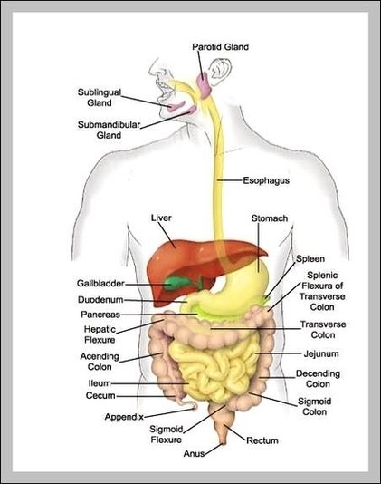
Physiology Of The Digestive System Image