

Anatomy Ear Image
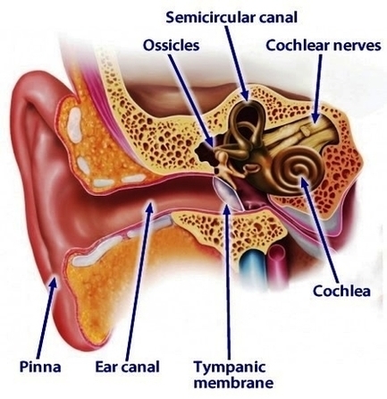
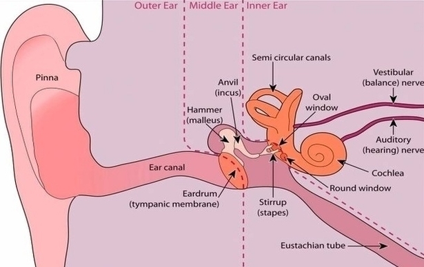
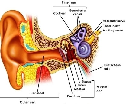
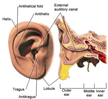
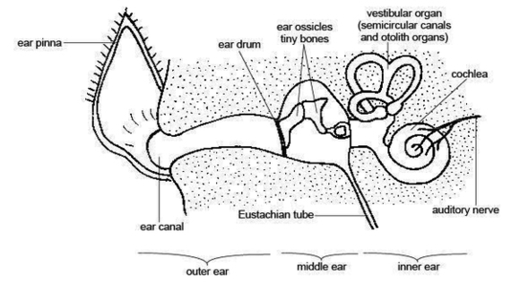
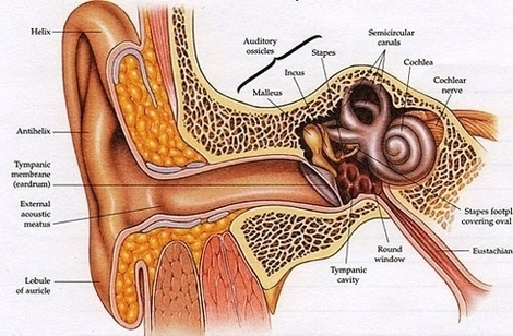
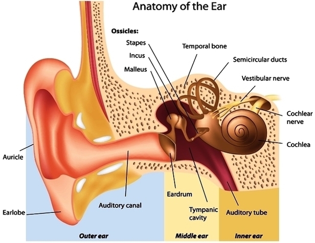
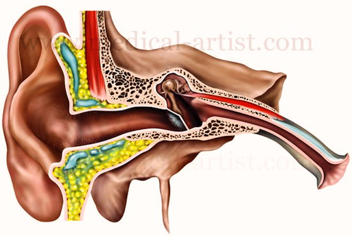
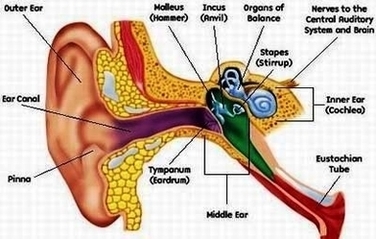
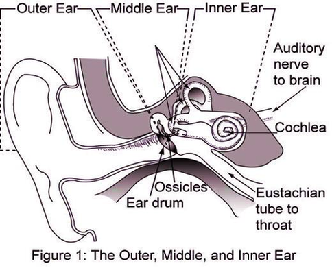
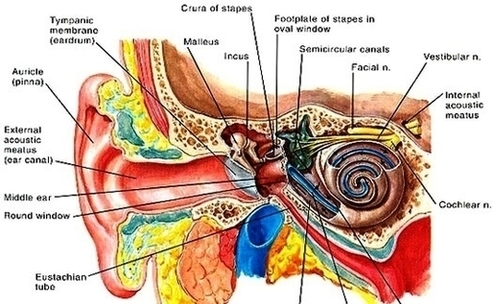
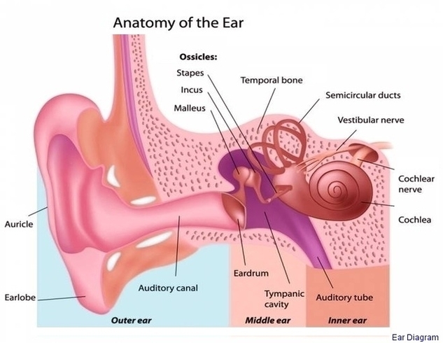
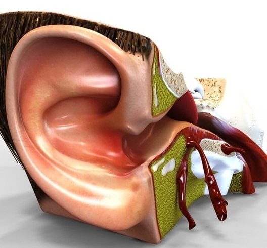
12,612 human ear anatomy stock photos, vectors, and illustrations are available royalty-free. Picture of the Ear. The spiral-shaped cochlea is part of the inner ear; it transforms sound into nerve impulses that travel to the brain. The fluid-filled semicircular canals View Diagram Anatomy Ear Image