Colorectal Cancer Small Image

4,035 colorectal cancer stock photos and images available, or search for colon cancer screening or colonoscopy to find more great stock photos and pictures. Doctor goes over a patient”s x-ray, screening for colon cancer. There is no single cause of colon cancer.

Colorectal cancer (CRC), bowel colon or rectal cancer. Abnormal growth of cells that invade or spread to other parts of the body. 3d render colorectal cancer stock pictures, royalty-free photos & images Internal view of the intestinal walls.

A colonoscopy is the most common test used to diagnose colorectal cancer. During a colonoscopy, the doctor looks inside the colon and rectum using a flexible tube with a light and lens on the end (called an endoscope).

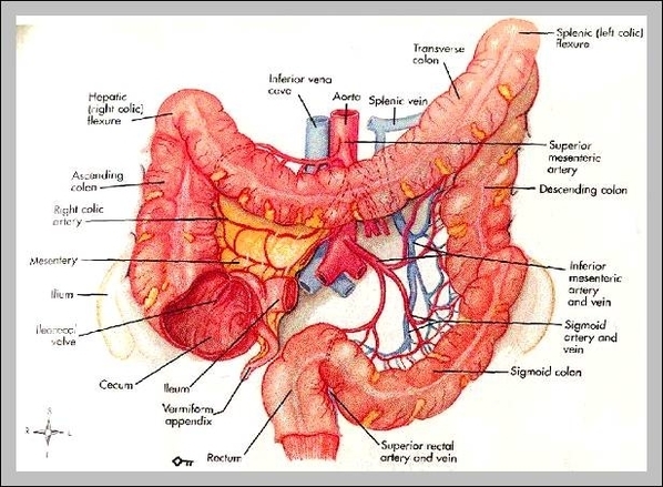
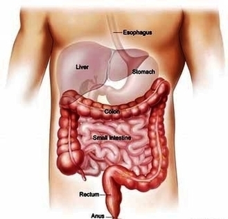
Colorectal Cancer Small Image Diagram - Chart - diagrams and charts with labels. This diagram depicts Colorectal Cancer Small Image
Colorectal Cancer Small Image

Cavity Tooth Decay Small Image

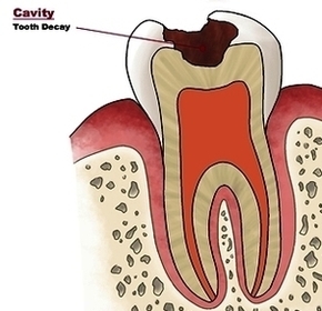
Cavities, also called tooth decay or caries, are caused by a combination of factors, including bacteria in your mouth, frequent snacking, sipping sugary drinks and not cleaning your teeth well. Cavities and tooth decay are among the world’s most common health problems. They’re especially common in children, teenagers and older adults.

49,828 tooth decay stock photos and images available, or search for tooth pain or bad teeth to find more great stock photos and pictures. Open mouth during drilling treatment at the dentist in dental clinic. Close-up.

The symptoms of tooth decay and cavities vary from child to child. Cavities don’t always cause symptoms. Sometimes children don’t know they have one until their dentist finds it. But your child may feel: Sensitivity to certain foods, such as sweets and hot or cold drinks

Cavity Tooth Decay Small Image Diagram - Chart - diagrams and charts with labels. This diagram depicts Cavity Tooth Decay Small Image

Cavity Tooth Decay Small Image

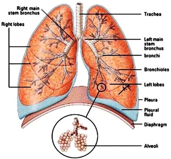
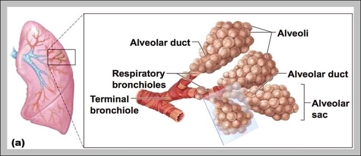
Lung Diagram Small Image

8,253 lung diagram stock photos, vectors, and illustrations are available royalty-free.

A healthy lung has a pinkish appearance, and if you could see it outside the body, it would look like a squishy, rubbery structure [4]. The lungs are located a little toward the posterior part of the human body, just below the collarbone, extending down to the diaphragm, the muscular partition that separates the chest and abdominal cavities.

What are Lungs: Definition. The spongy air-filled conical organs occupying most of the thoracic (chest) cavity in humans are known as the lungs [1]. It is one of the primary respiratory organs where the gas exchange occurs after the inhaled air enters the lungs via the trachea, through the bronchi and bronchioles [2].

Lung Diagram Small Image Diagram - Chart - diagrams and charts with labels. This diagram depicts Lung Diagram Small Image

Lung Diagram Small Image

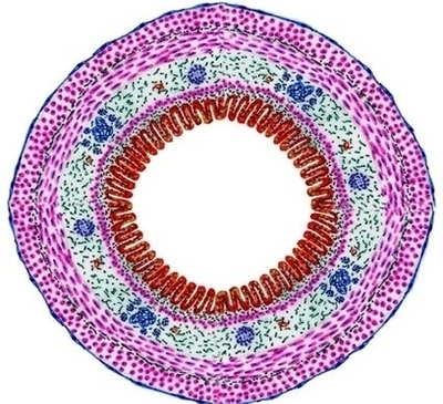
Tubular Organ Diagram Small Image

You know esophagus is the best organ that have all the layers of a tubular organ. I am going to describe the different layers from esophagus histology slide. The mucosa of esophagus consists of lamina epithelium, lamina propria and lamina muscularis.

5,814 male human anatomy diagram stock photos and images available, or start a new search to explore more stock photos and images. Male silhouette (contour) on white background, vector. Male, female and children’s silhouette on white background,…

The microscopic images of the urinary bladder and the labeled diagram might help you easily identify the slide under the light microscope. Most characteristic features of the urinary bladder slide are found in the tunica mucosa layer, lining epithelium, and tunica muscularis layer.

Tubular Organ Diagram Small Image Diagram - Chart - diagrams and charts with labels. This diagram depicts Tubular Organ Diagram Small Image

Tubular Organ Diagram Small Image

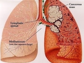
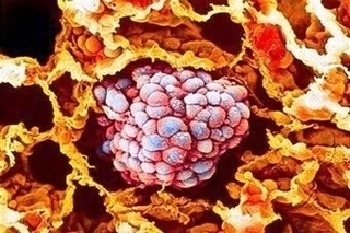
Small Cell Lung Cancer Treatment Image

Tests and procedures that examine the lungs are used to diagnose and stage small cell lung cancer. Certain factors affect prognosis (chance of recovery) and treatment options. For most patients with small cell lung current treatments do not cure the cancer.

For practical reasons, small cell lung cancer (SCLC) is usually staged as either limited or extensive. In most cases, SCLC has already spread by the time it is found, so chemotherapy (chemo) is usually part of treatment.

Radiation uses high-energy rays (such as x-rays) to kill cancer cells. In limited stage small cell lung cancer it’s most often used along with chemo to treat the tumor and lymph nodes in the chest. Or it could be used on the brain to try to keep the cancer from spreading there.

Small Cell Lung Cancer Treatment Image Diagram - Chart - diagrams and charts with labels. This diagram depicts Small Cell Lung Cancer Treatment Image

Small Cell Lung Cancer Treatment Image

Parts Of Small Intestine

Parts Of Small Intestine Diagram - Chart - diagrams and charts with labels. This diagram depicts Parts Of Small Intestine

parts of small intestine