Colorectal Cancer Lg Image

4,035 colorectal cancer stock photos and images available, or search for colon cancer screening or colonoscopy to find more great stock photos and pictures. Doctor goes over a patient”s x-ray, screening for colon cancer. There is no single cause of colon cancer.

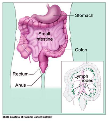
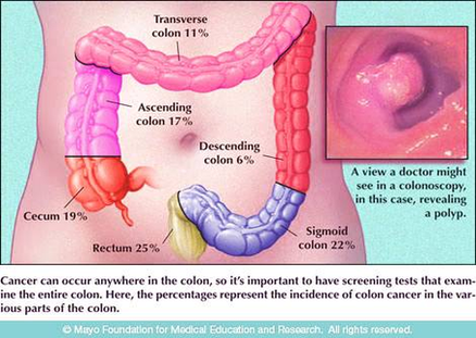
Colorectal cancer starts in the cells of the colon or rectum. A cancerous (malignant) tumour is a group of cancer cells that can grow into nearby tissue and destroy it. The tumour can also spread (metastasize) to other parts of the body.

But in some cases, changes to colon or rectal cells can cause colorectal cancer. Most often, colorectal cancer starts in gland cells that line the wall of the colon or rectum.

Colorectal Cancer Lg Image Diagram - Chart - diagrams and charts with labels. This diagram depicts Colorectal Cancer Lg Image
Colorectal Cancer Lg Image

Colorectal Cancer Lg Image

4,035 colorectal cancer stock photos and images available, or search for colon cancer screening or colonoscopy to find more great stock photos and pictures. Doctor goes over a patient”s x-ray, screening for colon cancer. There is no single cause of colon cancer.

Colorectal cancer starts in the cells of the colon or rectum. A cancerous (malignant) tumour is a group of cancer cells that can grow into nearby tissue and destroy it. The tumour can also spread (metastasize) to other parts of the body.

But in some cases, changes to colon or rectal cells can cause colorectal cancer. Most often, colorectal cancer starts in gland cells that line the wall of the colon or rectum.

Colorectal Cancer Lg Image Diagram - Chart - diagrams and charts with labels. This diagram depicts Colorectal Cancer Lg Image

Colorectal Cancer Lg Image

Peptic Ulcers Lg Enlg Image

210 stomach ulcer stock photos and images available, or search for stomach ulcer icon or human stomach ulcer to find more great stock photos and pictures. Gastric Ulcer, X-Ray.

A peptic ulcer of the stomach is called a gastric ulcer, an ulcer of the duodenum is a duodenal ulcer, and a peptic ulcer of the esophagus is an esophageal ulcer. A peptic ulcer occurs when the lining of these organs is corroded by the acidic digestive juices which are secreted by the stomach cells.

A peptic ulcer is a lesion in the tissue of the upper part of the gastrointestinal tract ( esophagus, stomach, duodenum). If it is located in the stomach, it’s called a gastric ulcer. A lesion in the esophagus is an esophageal ulcer.

Peptic Ulcers Lg Enlg Image Diagram - Chart - diagrams and charts with labels. This diagram depicts Peptic Ulcers Lg Enlg Image

Peptic Ulcers Lg Enlg Image

Nutrition Label Tricks Lg Image

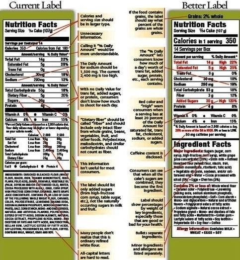
Nutrition facts show quantities of ingredients in products, including calories, fat, saturated fat, sodium, sugar, protein, carbohydrates, fiber and vitamins nutrition label stock pictures, royalty-free photos & images Nutrition label collage of multiple packaging labels.

Step 1. Which version of our Nutrition Label Generator would you like to use? Step 2. Choose a Nutrition Label Format The most basic nutrition label. Vertical layout. Standard nutrition label with extra fields for misc. vitamins. Vertical layout. Tabbed nutrition label for bottles & round containers. Horizontal layout. Step 3.

NOTE: FDA has issued final changes to update the Nutrition Facts label for packaged foods. For more information, see Changes to the Nutrition Facts Label. (For educational purposes only. These labels do not meet the labeling requirements described in 21 CFR 101.9.)

Nutrition Label Tricks Lg Image Diagram - Chart - diagrams and charts with labels. This diagram depicts Nutrition Label Tricks Lg Image

Nutrition Label Tricks Lg Image

Colon Lg Caption Figure Image

The following is an example of a caption inside a figure environment: begin{ figure } [h!] caption{ A picture of the universe! } includegraphics[width=0.5 extwidth]{ universe } end{ figure } The caption may be put either before or after the image in the figure.

We can simply add the following command to the figure environment: We do not need to include the figure number inside the caption, because LaTeX automatically keeps track of the numbering of the figures, and it’ll display it while displaying the caption.

We can simply add the following command to the figure environment: We do not need to include the figure number inside the caption, because LaTeX automatically keeps track of the numbering of the figures, and it’ll display it while displaying the caption. The following is an example of a caption inside a figure environment:

Colon Lg Caption Figure Image Diagram - Chart - diagrams and charts with labels. This diagram depicts Colon Lg Caption Figure Image

Colon Lg Caption Figure Image

Colon Lg Caption Image

The caption image tool is only one of the many features the editor offers. Whether you are a non-designer, a business owner, or a teacher, you can use Pixelied to create stunning visuals from scratch or based on templates. Pixelied is worth its weight in gold.

– GeeksforGeeks How to set caption for an image using HTML ? The tag in HTML is used to set a caption to the figure element in a document. This tag is new in HTML5. How to define an image caption?

Emphasize your caption. Make your caption hover above the image with well-placed shadows. Fit the caption to your design. Adjust the line height, so your caption fits your visual perfectly. Deliver a clear message.

Colon Lg Caption Image Diagram - Chart - diagrams and charts with labels. This diagram depicts Colon Lg Caption Image

Colon Lg Caption Image

Colon Lg Image

The colon, which is another name for the large intestine, is an important part of the digestive system.

Once or twice per day, when the body is ready for a bowel movement, the waste is dumped into the rectum. There are many diseases and conditions that are associated with the colon. Colorectal cancer is cancer that occurs in the colon or in the lower colon near the rectum. It is one of the most common causes of cancer-associated death.

The colon is comprised of four layers of tissue, similar to other regions of the digestive tract. These include: Mucosa: This is the innermost layer and is made of simple columnar epithelial tissue, making it smooth (compared to the small intestine, which contains villi, small fingerlike protrusions).

Colon Lg Image Diagram - Chart - diagrams and charts with labels. This diagram depicts Colon Lg Image

Colon Lg Image