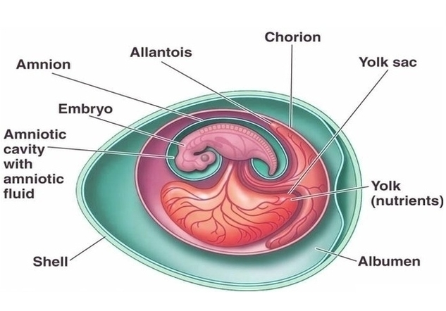
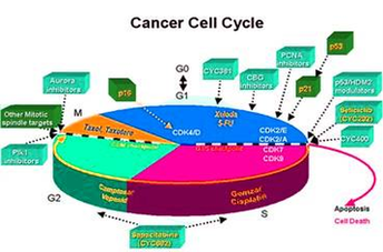
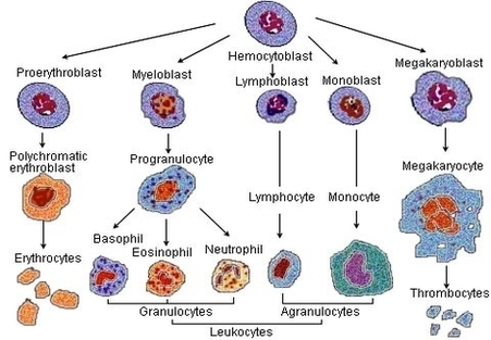
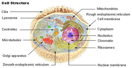
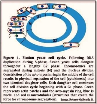
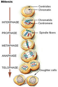
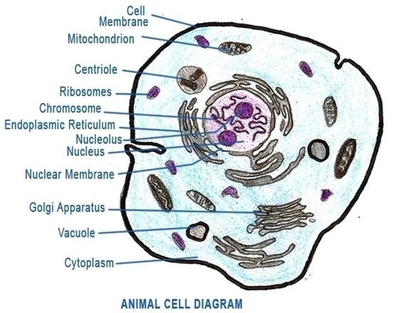
Cancer Cell Diagram Image
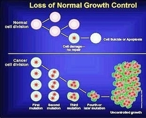
4,046 cancer cell stock photos and images available, or search for cancer patient or cancer research to find more great stock photos and pictures. Cancer cells appear through a series of genetic and epigenetic changes. Some of these changes may View Diagram Cancer Cell Diagram Image