Asthma Airways Image

This human anatomy diagram with labels depicts and explains the details and or parts of the Asthma Airways Image. Human anatomy diagrams and charts show internal organs, body systems, cells, conditions, sickness and symptoms information and/or tips to ensure one lives in good health.

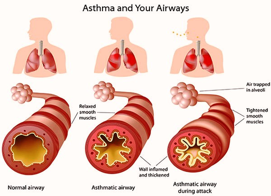
In normal breathing, the airways to the lungs are completely open. Air moves freely in and out. But in someone who lives with asthma, these airways swell and become inflamed. As the airways grow irritated and sensitive, they start to react to various asthma triggers, which are things that you are exposed to every day. 2

Asthmatic Airway Cells include lung fibroblasts, bronchial epithelial and bronchial smooth muscle cells. Obstructed bronchial tube illustration representing asthma. 3D illustration. Salamol (Salbutamol Sulphate) inhaler for relief of broncheal spasms and inflammation.

Besides having trouble breathing, a person with asthma experiences coughing and shortness of breath. You can feel as if something is tightening around your chest. You may make a whistling sound called wheezing as you inhale and exhale. 4

Asthma Airways Image

Asthma Airways Image


Tags: ,