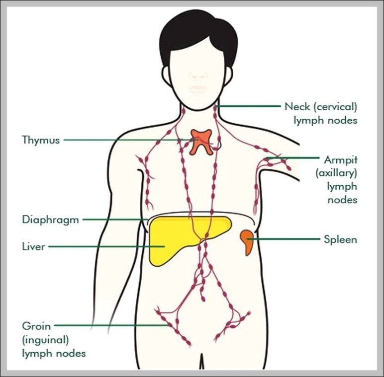
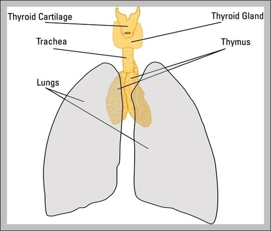
Thymus Diagram Image
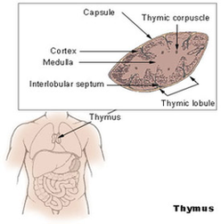
The gland extends from as high as the lower border of the thyroid gland to the fourth costal cartilage downwards. The thymus is of a pinkish-grey color, soft, and lobulated on its surfaces. Embryologically it is derived from the third View Diagram Thymus Diagram Image