Spinal Vertebrae Chart

This human anatomy diagram with labels depicts and explains the details and or parts of the Spinal Vertebrae Chart. Human anatomy diagrams and charts show internal organs, body systems, cells, conditions, sickness and symptoms information and/or tips to ensure one lives in good health.

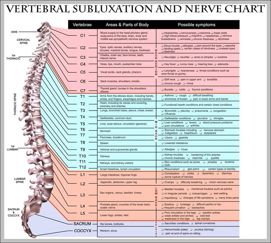
These regions are called the cervical spine, thoracic spine, lumbar spine, sacrum and coccyx. There are seven cervical vertebrae, twelve thoracic vertebrae and five lumbar vertebrae. The number of vertebrae in a region can vary but overall the number remains the same.

The vertebral arch is posterior, meaning it faces the back of a person. Together, these enclose the vertebral foramen, which contains the spinal cord. Because the spinal cord ends in the lumbar spine, and the sacrum and coccyx are fused, they do not contain a central foramen.

The second cervical vertebra, C2, is also known as the “axis” because it allows the skull and atlas to rotate to the left and right. Thoracic: The 12 vertebrae in the chest region form the spine’s thoracic region. Thoracic vertebrae are larger and stronger than cervical vertebrae but are much less flexible.

Spinal Vertebrae Chart

spinal vertebrae chart


Tags: , , , , , , ,