Cyclacel Cell Cycle Diagram1 Image

This human anatomy diagram with labels depicts and explains the details and or parts of the Cyclacel Cell Cycle Diagram1 Image. Human anatomy diagrams and charts show internal organs, body systems, cells, conditions, sickness and symptoms information and/or tips to ensure one lives in good health.

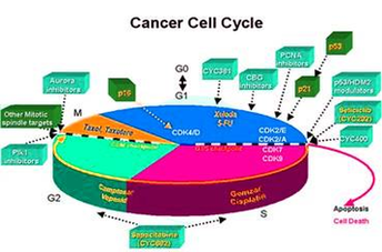
The phases are: 1. G1 (gap1) phase 2. S (synthesis) phase 3. G2 (gap 2) phase 4. M (mitosis) phase. Cell Cycle: Phase # 1.

Our strategy is to build a diversified portfolio of novel drug candidates to address areas of high unmet medical needs. Cyclacel is a pioneer in the field of cell cycle biology. Our vision is to improve patient healthcare by translating cancer biology into medicines.

The G 1 phase is set in immediately after the cell division. It is characterised by a change in the chromosome from the condensed mitotic state to the more extended interphase state and by a series of metabolic events leading to initiation of DNA replication. G, phase of cell cycle varies in length from cell to cell within the same cell population.

Cyclacel Cell Cycle Diagram1 Image

Cyclacel Cell Cycle Diagram1 Image


Tags: , , ,