Skip to content

Human Anatomy Diagrams

Menu

Primary menu

  • Top
  • Anatomy
  • Muscles
  • Systems
  • Body Parts
  • Organs
  • Cells
  • Bones
  • Teeth
  • Cancer
  • Health

Author Archives: anatomy

Diagram Of Tooth Implant Procedure Image

Posted on June 22, 2022 by anatomy
Diagram Of Tooth Implant Procedure Image

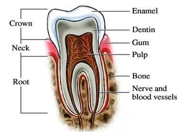
To understand how a dental implant procedure is performed, you should know the parts of the dental implant. A conventional dental implant consists of 3 parts: the implant body, abutment, and dental prosthesis. The implant body: it is similar in View Diagram Diagram Of Tooth Implant Procedure Image→

Posted in Anatomy | Tagged implant, procedure, tooth

Anatomy Of The Human Eye Cross Section View Image

Posted on June 22, 2022 by anatomy
Anatomy Of The Human Eye Cross Section View Image

Eye – Cross Section. A human eyeball cross section showing the following structures: iris, anterior limiting membrane, posterior limiting membrane, tendon of Eye anatomy, Human eye cross section physiology, Model of cornea and lens for ophthalmologist. Eye anatomy, Human eye View Diagram Anatomy Of The Human Eye Cross Section View Image→

Posted in Anatomy | Tagged cross, eye, section, view

How To Become A Nurse Image

Posted on June 22, 2022 by anatomy

How To Become A Nurse Image

Posted in Anatomy | Tagged a, become, how, nurse, to

Stages Of Periodontitis Image

Posted on June 22, 2022 by anatomy
Stages Of Periodontitis Image

Periodontal disease is broken up into four separate stages: gingivitis, slight periodontal disease, moderate periodontal disease, and advanced periodontal disease. Gingivitis. Gingivitis is the only stage of periodontal disease that is reversible as it has not yet had time to View Diagram Stages Of Periodontitis Image→

Posted in Anatomy | Tagged periodontitis, stages

Human Body Muscle Diagram Image

Posted on June 22, 2022 by anatomy
Human Body Muscle Diagram Image

43,556 human body diagram stock photos, vectors, and illustrations are available royalty-free. 128,412 muscle anatomy stock photos, vectors, and illustrations are available royalty-free. Last Updated: Jul 16, 2019 The muscular system is responsible for the movement of the human body. View Diagram Human Body Muscle Diagram Image→

Posted in Anatomy, Muscles | Tagged body, human, muscle

Diabetes Diet Food Tips Banner Larges Image

Posted on June 22, 2022 by anatomy
Diabetes Diet Food Tips Banner Larges Image

So an effort to use charts will be done. The way to get the optimal version of the diabetic diet chart is to consistently do every element in it. If you have done it regularly and don’t try to cheat View Diagram Diabetes Diet Food Tips Banner Larges Image→

Posted in Anatomy | Tagged banner, diabetes, diet, food, larges, tips

Retina Layers Diagram Image

Posted on June 22, 2022 by anatomy
Retina Layers Diagram Image

Layers of the Retina. Choroid – This is made up of a layer of blood vessels that supply oxygen and nutrients to the retina. Defect in the CHM gene can cause choroideremia, leaky blood vessels can expand in the retina View Diagram Retina Layers Diagram Image→

Posted in Cells | Tagged layers, retina

What Is Blood Image

Posted on June 22, 2022 by anatomy
What Is Blood Image

Picture of Blood 1 • Red blood cells, which carry oxygen to the tissues 2 • White blood cells, which fight infections 3 • Platelets, smaller cells that help blood to clot More … 1,418,380 blood stock photos, vectors, and View Diagram What Is Blood Image→

Posted in Cells | Tagged blood, Is, What

Pregnancy Weeks Fetus Development Image

Posted on June 22, 2022 by anatomy
Pregnancy Weeks Fetus Development Image

Baby’s Development at 20 Weeks. At 20 weeks, the developing fetus is about 6 inches long (13.2 cm) and may weigh about 10 ounces. The baby may begin to make movements that the mother can feel at about 19 to View Diagram Pregnancy Weeks Fetus Development Image→

Posted in Cells | Tagged development, fetus, pregnancy, weeks

Human Body Muscle System Image

Posted on June 22, 2022 by anatomy
Human Body Muscle System Image

105,188 human muscle anatomy stock photos, vectors, and illustrations are available royalty-free. Last Updated: Jul 16, 2019 The muscular system is responsible for the movement of the human body. Attached to the bones of the skeletal system are about 700 View Diagram Human Body Muscle System Image→

Posted in Muscles, System | Tagged body, human, muscle, system

Bones In Buttocks Image

Posted on June 22, 2022 by anatomy
Bones In Buttocks Image

577 pics of the female buttocks anatomy stock photos and images available, or start a new search to explore more stock photos and images. Houman body parts flat line icons set. Bone Structure. The pelvis is the part of your View Diagram Bones In Buttocks Image→

Posted in Anatomy, Bones | Tagged bones, buttocks

Diabetes Mellitus Type Image

Posted on June 22, 2022 by anatomy
Diabetes Mellitus Type Image

9,868 diabetes mellitus stock photos, vectors, and illustrations are available royalty-free. Type 1 and Type 2 Diabetes Mellitus. Insulin-Dependent Diabetes Mellitus and Non Insulin-Dependent Diabetes Mellitus. Insulin resistance and insufficient insulin production. Medical doctor drawing diabetes on the virtual screen. View Diagram Diabetes Mellitus Type Image→

Posted in Anatomy | Tagged diabetes, mellitus

Diagram First Trimester Pregnancy Stages Image

Posted on June 22, 2022 by anatomy
Diagram First Trimester Pregnancy Stages Image

A typical pregnancy lasts 40 weeks from the first day of your last menstrual period (LMP) to the birth of the baby. It is divided into three stages, called trimesters: first trimester, second trimester, and third trimester. The fetus undergoes View Diagram Diagram First Trimester Pregnancy Stages Image→

Posted in Anatomy | Tagged First, pregnancy, stages, trimester

Shoulder Xray Acj Normaltures Image

Posted on June 22, 2022 by anatomy
Shoulder Xray Acj Normaltures Image

Normal radiographic measurements of the shoulderare important in the evaluation of the osseous relationships in plain radiography. Normal measurements do not rule out pathology and must be considered in the context of other findings and the clinical presentation. acromioclavicular (AC) View Diagram Shoulder Xray Acj Normaltures Image→

Posted in Anatomy | Tagged acj, normaltures, shoulder, xray

Embryo Fetus Image

Posted on June 22, 2022 by anatomy

Embryo Fetus Image

Posted in Anatomy | Tagged embryo, fetus

Reproductive Male Diagram Image

Posted on June 22, 2022 by anatomy
Reproductive Male Diagram Image

The male reproductive system primarily consists of the paired testes and the penis. In addition accessory sex glands contribute to seminal fluid. The male reproductive system matures during adolescence and remains active for the remainder of the lifespan of the View Diagram Reproductive Male Diagram Image→

Posted in Cells | Tagged male, reproductive

Tooth Anatomy Image

Posted on June 22, 2022 by anatomy
Tooth Anatomy Image

WebMD’s Teeth Anatomy Page provides a detailed diagram and definition of the teeth, inlcuding types, names, and parts of the teeth. Skip to main content • Pulp: The softer, living inner structure of teeth. Blood vessels and nerves run through View Diagram Tooth Anatomy Image→

Posted in Anatomy | Tagged tooth

Dry Eye Label Image

Posted on June 22, 2022 by anatomy
Dry Eye Label Image

These are thick-rimmed glasses that wrap around your head in order to reduce your eyes’ exposure to fog and external moisture, thus limiting your susceptibility to the symptoms of dry eye syndrome. According to the former-microbiologist-turned-health-journalist, Bryn Nelson, dry eye View Diagram Dry Eye Label Image→

Posted in Anatomy | Tagged dry, eye, label

Labeled Skeleton Diagram Image

Posted on June 22, 2022 by anatomy
Labeled Skeleton Diagram Image

62 labeled diagram of the skeletal system pic stock photos and images available, or start a new search to explore more stock photos and images. Skeletal Poster Human skeletal system poster containing detailed information about the skeletal structure. Blank skeleton View Diagram Labeled Skeleton Diagram Image→

Posted in Anatomy | Tagged labeled, skeleton

Diagram Illu Synovial Joint Image

Posted on June 22, 2022 by anatomy
Diagram Illu Synovial Joint Image

The structure of a synovial joint is demonstrated by a diagram in which the articulating bones are surrounded by the articular capsule, which comprises an exterior fibrous capsule and an interior synovial membrane. Start studying label the synovial joint. Learn View Diagram Diagram Illu Synovial Joint Image→

Posted in Anatomy | Tagged illu, joint, synovial

Post navigation

← Older posts
Newer posts →

Primary Sidebar Widget Area

Top Diagrams

  • Human Body Organ Locations Human Body Organ Locations

  • Map Of Body Muscles Map Of Body Muscles

  • Spinal Vertebrae Chart Spinal Vertebrae Chart

  • Human Organs Back View Human Organs Back View

  • Female Abdominal Anatomy Female Abdominal Anatomy

  • Male Body Organs Diagram Male Body Organs Diagram

  • Map Of Human Organs Map Of Human Organs

  • Female Muscular System Female Muscular System

  • Lower Back Hip Anatomy Lower Back Hip Anatomy

  • Picture Of Organs On Left Side Of Body Picture Of Organs On Left Side Of Body

  • Male Anatomy Pictures Male Anatomy Pictures

  • Female Human Anatomy Organs Female Human Anatomy Organs

  • Lower Back Anatomy Pictures Lower Back Anatomy Pictures

  • Muscular System Full Body Diagram Muscular System Full Body Diagram

  • Groin Muscles Anatomy Groin Muscles Anatomy

Latest Posts

  • Cutaneous Innervation to the Gluteal Region
  • Submental Triangle of the Neck
  • Diagram of the Descent of the Testis Embryology
  • Pancreatic duct Bile duct and Major duodenal Papilla
  • Direct Inguinal Hernia vs Indirect Inguinal Hernia Regional Anatomy
  • Muscles of the Medial Thigh Hip Adductors
  • Overview of the Mesentery
  • Aortic Aneurysm
  • Great Vessels of the Heart
  • Inferior Alveolar Nerve Course Supply
  • Sagittal View of the Subdivisions of the Peritoneum
  • Ligaments of the Elbow Joint Lateral and Medial Aspect
  • Borders of the Femoral Triangle
  • Palmar Fascia of the Hand
  • Knee 3D

Categories

  • Anatomy
  • Body Parts
  • Bones
  • Cancer
  • Cells
  • Health
  • Muscles
  • Organs
  • System
  • Teeth
  • Top

Top Tags

  • muscles (205)
  • muscle (171)
  • human (156)
  • human muscles (138)
  • muscles diagram (138)
  • muscles anatomy (138)
  • human muscle diagram (136)
  • muscular system (136)
  • body (119)
  • system (119)
  • body organs (119)
  • blood (82)
  • parts of a human body (77)
  • human body parts (77)
  • body parts (77)
  • organs (67)
  • organ (67)
  • pregnancy (66)
  • diabetes (62)
  • organ anatomy (62)
Copyright © 2026 . All Rights Reserved.
Theme: Catch Box by Catch Themes
Scroll Up