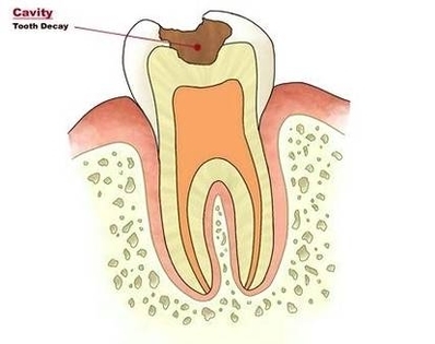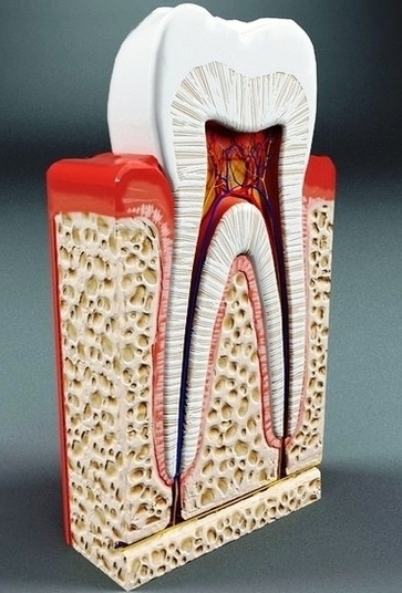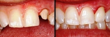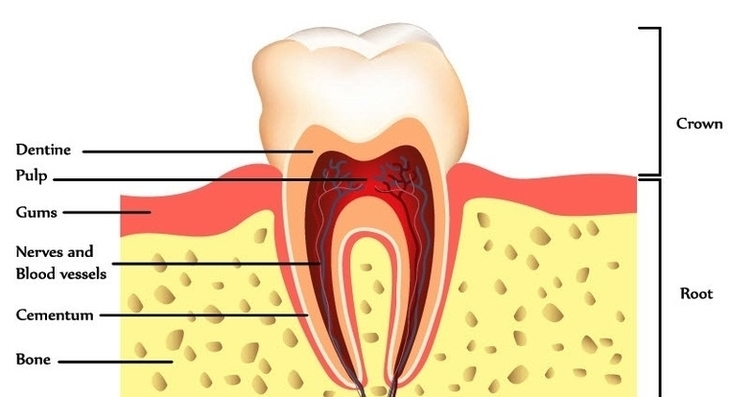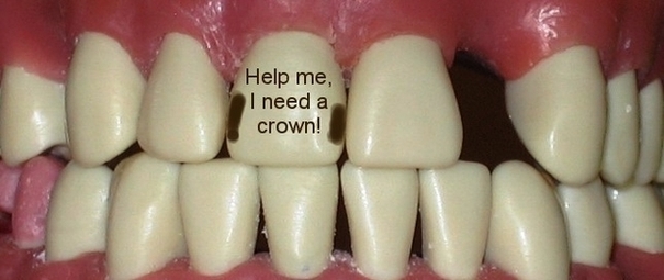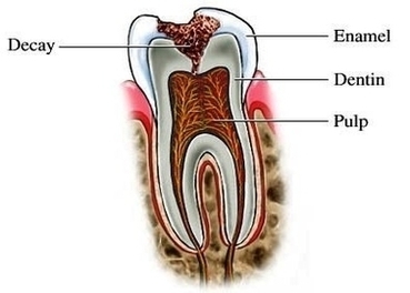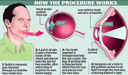

Cavity Tooth Decay Small Image
Cavities, also called tooth decay or caries, are caused by a combination of factors, including bacteria in your mouth, frequent snacking, sipping sugary drinks and not cleaning your teeth well. Cavities and tooth decay are among the world’s most common View Diagram Cavity Tooth Decay Small Image