The Human Eye And Internal Anatomy Of The Eyeball Images Image
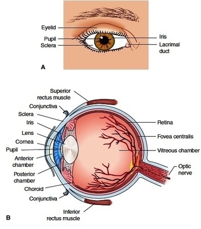
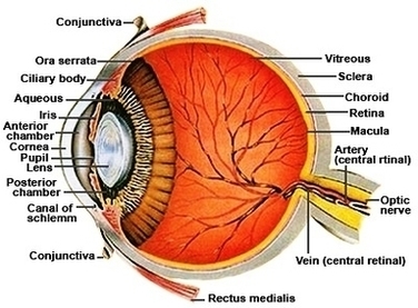
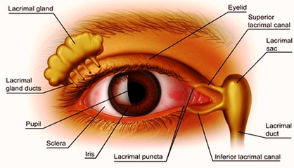
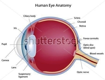
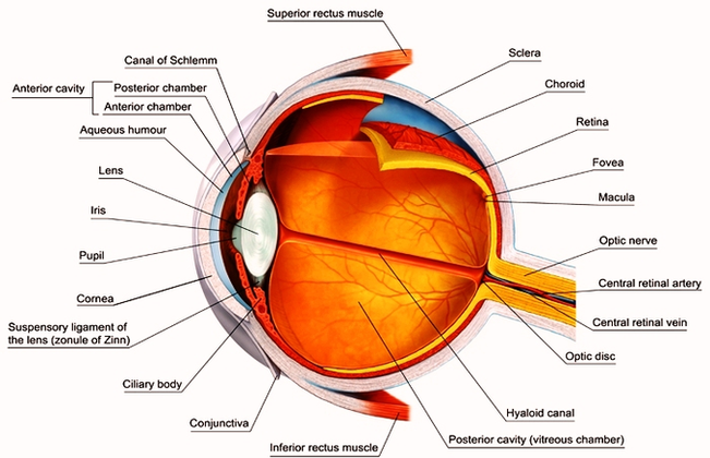
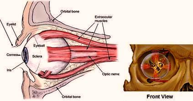
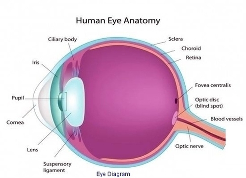
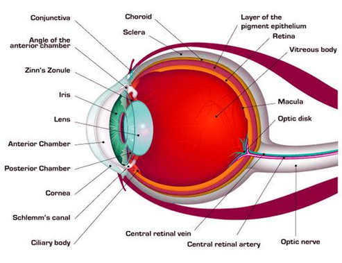
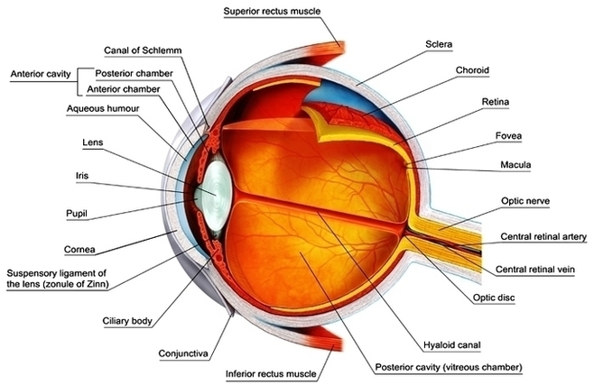
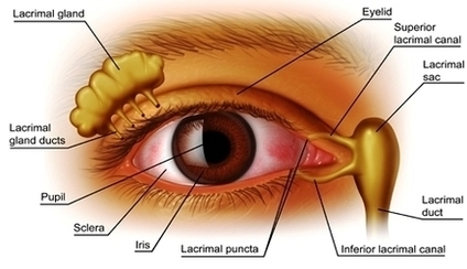
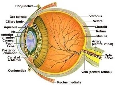
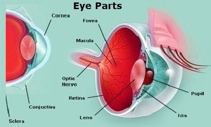
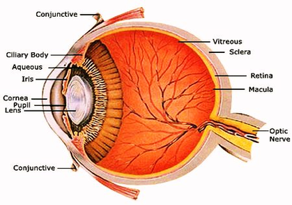
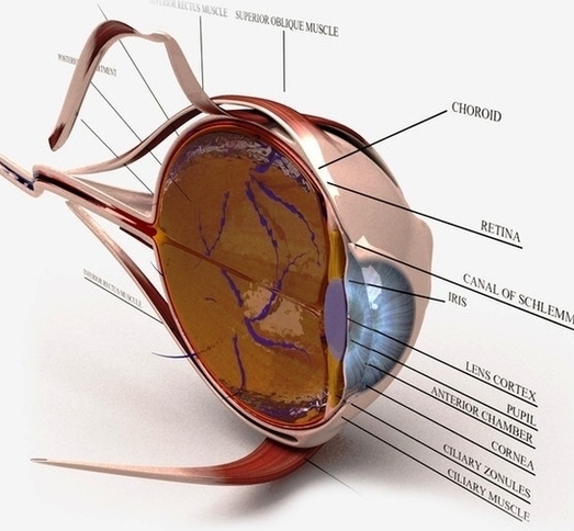
Picture of Eye Anatomy Detail The eye is our organ of sight. The eye has a number of components which include but are not limited to the cornea, iris, pupil, lens, retina, macula, optic nerve, choroid and vitreous. Cornea: clear View Diagram The Human Eye And Internal Anatomy Of The Eyeball Images Image