Diagram Illu Synovial Joint Image

This human anatomy diagram with labels depicts and explains the details and or parts of the Diagram Illu Synovial Joint Image. Human anatomy diagrams and charts show internal organs, body systems, cells, conditions, sickness and symptoms information and/or tips to ensure one lives in good health.

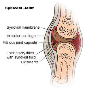
The structure of a synovial joint is demonstrated by a diagram in which the articulating bones are surrounded by the articular capsule, which comprises an exterior fibrous capsule and an interior synovial membrane. Start studying label the synovial joint. Learn vocabulary, terms, and more with flashcards, games, and other study tools.

Start studying label the synovial joint. Learn vocabulary, terms, and more with flashcards, games, and other study tools. Labeled Diagram of the Knee Joint Knee joint is one of the most important hinge joints of our body. Its complexity and its efficiency is the best example of God’s creation.

Synovial joints are directly supported by ligaments, which span between the bones of the joint. These may be located outside of the articular capsule (extrinsic ligaments), incorporated or fused to the wall of the articular capsule (intrinsic ligaments), or found inside of the articular capsule (intracapsular ligaments).

Diagram Illu Synovial Joint Image

Diagram Illu Synovial Joint Image


Tags: , ,