
Tag Archives: synovial
Synovial Joint Diagram Image
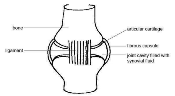
The structure of a synovial joint is demonstrated by a diagram in which the articulating bones are surrounded by the articular capsule, which comprises an exterior fibrous capsule and an interior synovial membrane. Start studying label the synovial joint. Learn View Diagram Synovial Joint Diagram Image
Diagram Illu Synovial Joint Image
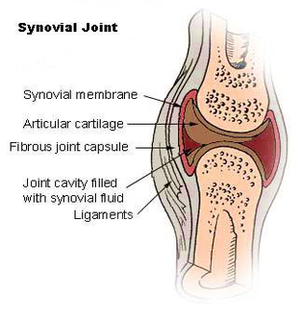
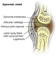
The structure of a synovial joint is demonstrated by a diagram in which the articulating bones are surrounded by the articular capsule, which comprises an exterior fibrous capsule and an interior synovial membrane. Start studying label the synovial joint. Learn View Diagram Diagram Illu Synovial Joint Image