Anatomy Of The Human Eye Cross Section View Image

This human anatomy diagram with labels depicts and explains the details and or parts of the Anatomy Of The Human Eye Cross Section View Image. Human anatomy diagrams and charts show internal organs, body systems, cells, conditions, sickness and symptoms information and/or tips to ensure one lives in good health.

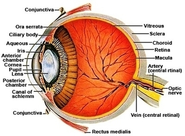
Eye – Cross Section. A human eyeball cross section showing the following structures: iris, anterior limiting membrane, posterior limiting membrane, tendon of Eye anatomy, Human eye cross section physiology, Model of cornea and lens for ophthalmologist. Eye anatomy, Human eye cross section physiology, Model of cornea Enlarged anatomical eye model.

A human eyeball cross section showing the following structures: iris, anterior limiting membrane, posterior limiting membrane, tendon of Eye anatomy, Human eye cross section physiology, Model of cornea and lens for ophthalmologist.

Cross section of vitreous anatomy. A, Cross section diagram of the eye with emphasis on the anatomical features of the vitreous. The vitreous is most firmly attached to the retina at the vitreous base, and it also has adhesions at the optic nerve, along vessels, at the fovea, and to the posterior lens capsule.

Anatomy Of The Human Eye Cross Section View Image

Anatomy Of The Human Eye Cross Section View Image


Tags: , , ,