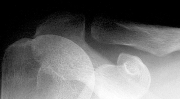
Shoulder Xray Acj Normaltures Image

This human anatomy diagram with labels depicts and explains the details and or parts of the Shoulder Xray Acj Normaltures Image. Human anatomy diagrams and charts show internal organs, body systems, cells, conditions, sickness and symptoms information and/or tips to ensure one lives in good health.

Normal radiographic measurements of the shoulderare important in the evaluation of the osseous relationships in plain radiography. Normal measurements do not rule out pathology and must be considered in the context of other findings and the clinical presentation. acromioclavicular (AC) joint space: 1-7 mm 4(narrower in the elderly)

Shoulder X-rays are common investigations in every Emergency Department, typically in the context of trauma, with shoulder dislocations being the most common pathology. An important distinction to note is what we mean when we request a “shoulder X-ray”. In most clinical scenarios this refers to a radiograph of the glenohumeral joint.

Standard acromioclavicular joint radiographs consist of a clavicle seriesincluding an AP and cephalic angled oblique (10-15º) views. Additional weight-bearing stress viewsmay be of benefit if: initial radiographs are normal, but an injury is suspected

Shoulder Xray Acj Normaltures Image

Shoulder Xray Acj Normaltures Image


Tags: , , ,