Diagram Illu Dige Tract Image

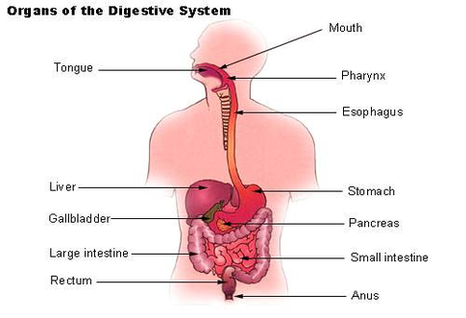
Looking at pictures of your GI tract can help you to pinpoint where symptoms such as abdominal pain may be coming from. This understanding can also help you to better describe your symptoms to your healthcare provider.

Terminal Ileum. The terminal ileum is the distal end of the small intestine that intersects with the large intestine.

The organs that make up the GI tract are the mouth, throat, esophagus, stomach, small intestine, large intestine, rectum, and anus. The GI tract is one part of the digestive system. 2

Diagram Illu Dige Tract Image Diagram - Chart - diagrams and charts with labels. This diagram depicts Diagram Illu Dige Tract Image

Diagram Illu Dige Tract Image

Diagram Illu Synovial Joint Image

The structure of a synovial joint is demonstrated by a diagram in which the articulating bones are surrounded by the articular capsule, which comprises an exterior fibrous capsule and an interior synovial membrane. Start studying label the synovial joint. Learn vocabulary, terms, and more with flashcards, games, and other study tools.

Start studying label the synovial joint. Learn vocabulary, terms, and more with flashcards, games, and other study tools. Labeled Diagram of the Knee Joint Knee joint is one of the most important hinge joints of our body. Its complexity and its efficiency is the best example of God’s creation.

Synovial joints are directly supported by ligaments, which span between the bones of the joint. These may be located outside of the articular capsule (extrinsic ligaments), incorporated or fused to the wall of the articular capsule (intrinsic ligaments), or found inside of the articular capsule (intracapsular ligaments).

Diagram Illu Synovial Joint Image Diagram - Chart - diagrams and charts with labels. This diagram depicts Diagram Illu Synovial Joint Image

Diagram Illu Synovial Joint Image

Diagram Of Illu Bone Growth Image

Bone growth diagrams show the progression of development of the bone over a period of time. This visually displays where a bone accepts blood vessels or where cartilage develops. This type of skeletal diagram also may show a cross section of a bone and the different layers within a bone: bone marrow, osteoclasts, cancellous bone, and cortical bone.

Figure 6.3.1 – Anatomy of a Long Bone: A typical long bone showing gross anatomical features. The wider section at each end of the bone is called the epiphysis (plural = epiphyses), which is filled internally with spongy bone, another type of osseous tissue.

Some skeletal diagrams show smaller sections of the human skeleton. These are usually a grouping, such as bones of the head, spine, feet and hands. There are many different types of skeletal diagrams.

Diagram Of Illu Bone Growth Image Diagram - Chart - diagrams and charts with labels. This diagram depicts Diagram Of Illu Bone Growth Image

Diagram Of Illu Bone Growth Image