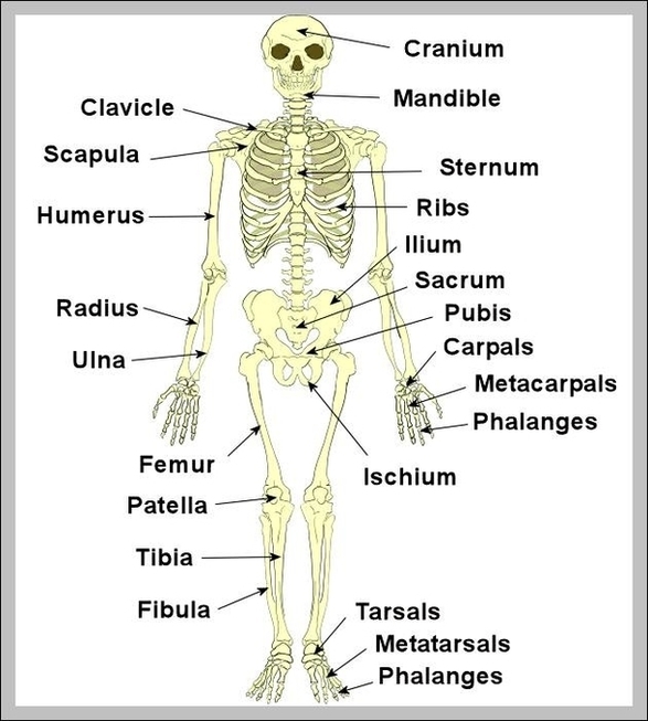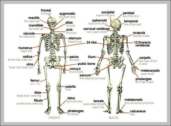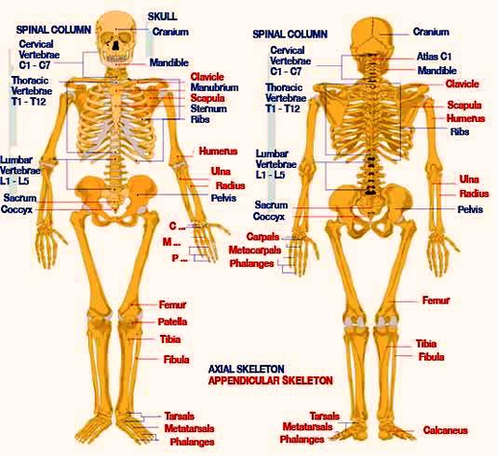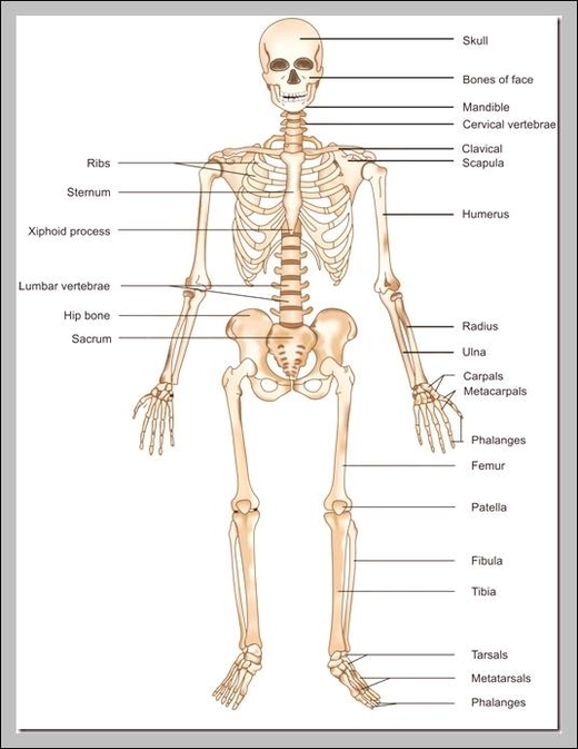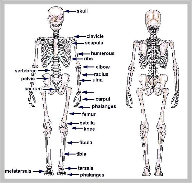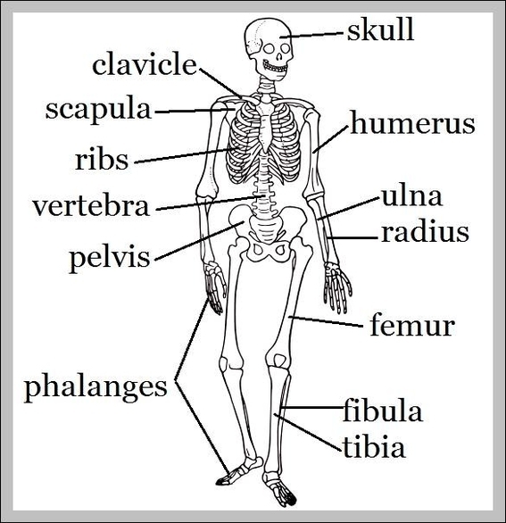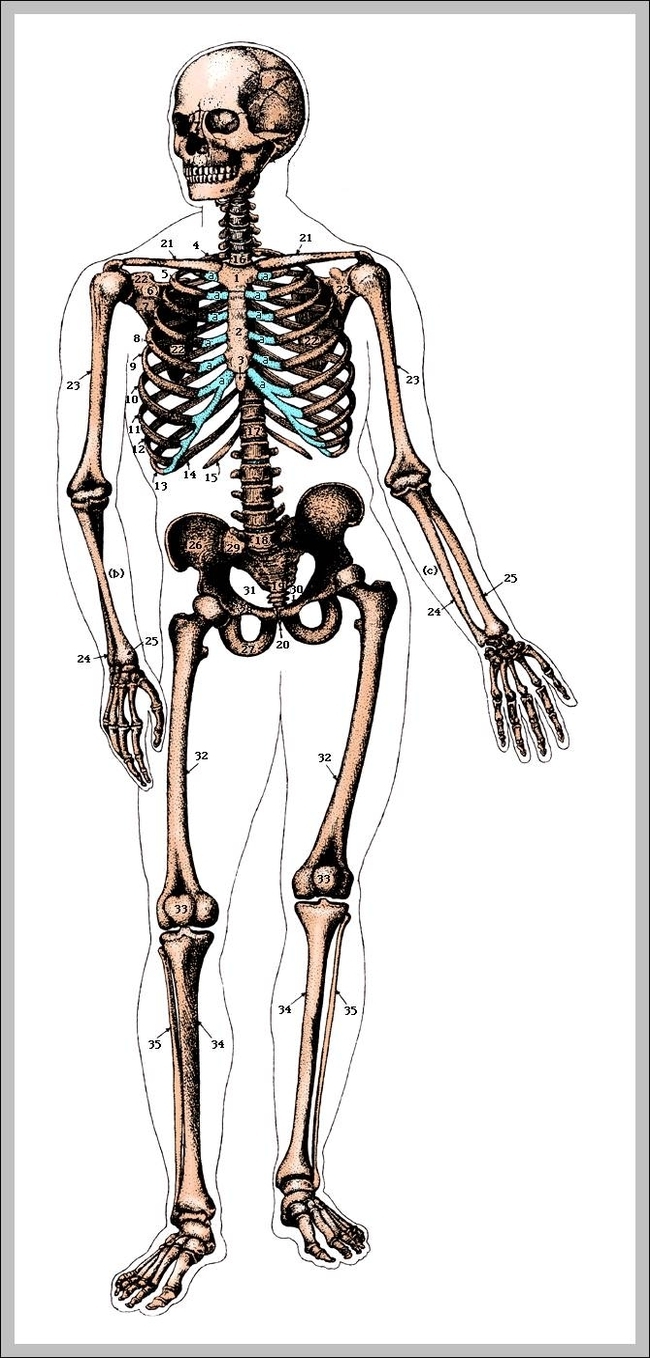
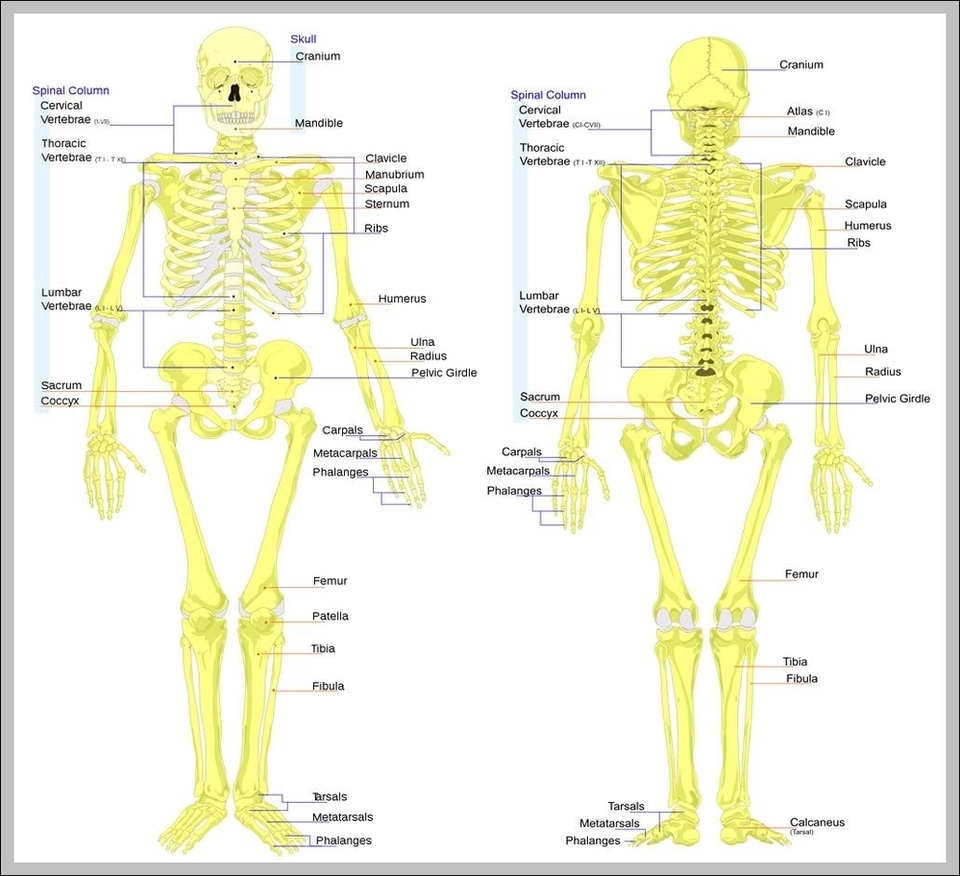
Labeled Skeleton Diagram Image
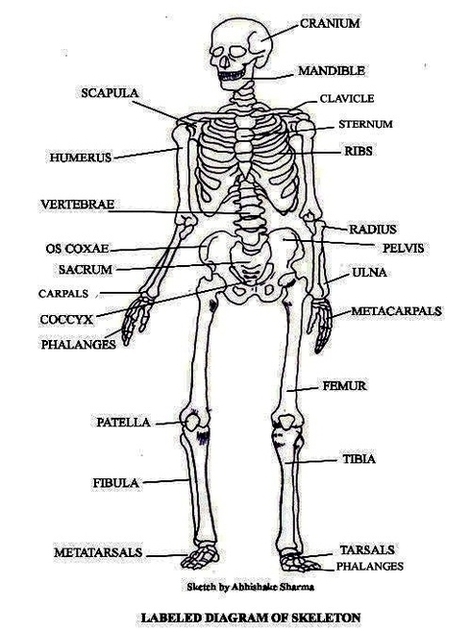
62 labeled diagram of the skeletal system pic stock photos and images available, or start a new search to explore more stock photos and images. Skeletal Poster Human skeletal system poster containing detailed information about the skeletal structure. Blank skeleton View Diagram Labeled Skeleton Diagram Image