Fixed Teeth Replacement Image
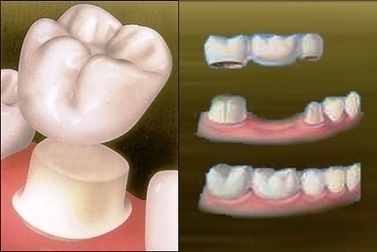
Fixed Bridge is second type of full arch tooth implant replacement. As the name suggest, your false teeth are permanent or non-removable. Only the dentist can remove the replacement teeth during your regular dental cleaning. Fixed Bridges are more expensive View Diagram Fixed Teeth Replacement Image