Knee Anatomy Diagram

This human anatomy diagram with labels depicts and explains the details and or parts of the Knee Anatomy Diagram. Human anatomy diagrams and charts show internal organs, body systems, cells, conditions, sickness and symptoms information and/or tips to ensure one lives in good health.

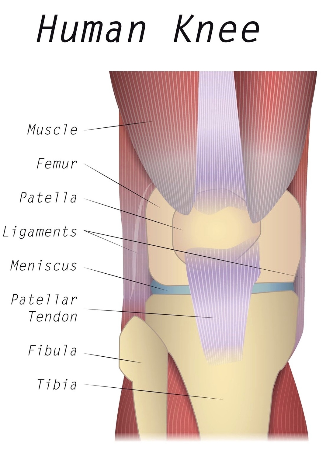
The most basic component of knee joint anatomy are the bones which provide the structure to the knee. There are four knee bones that fit together to make two different knee joints:

Our interactive 3D knee diagram is an informative 360 degree rotating model. Each of the 6 sections ( Bones, Connective Tissue 1, Connective Tissue 2, Deep Muscles, Muscles & Skin) can be opened up, rotated left or right and viewed more closely.

To understand one of the most complex joints of our body i.e. the knee joint, you need a perfectly labeled diagram of the knee. This will help you to understand the mechanism as well as the working.

Knee Anatomy Diagram

Knee anatomy diagram


Tags: , , ,