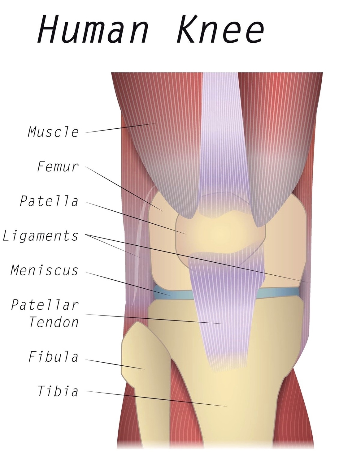
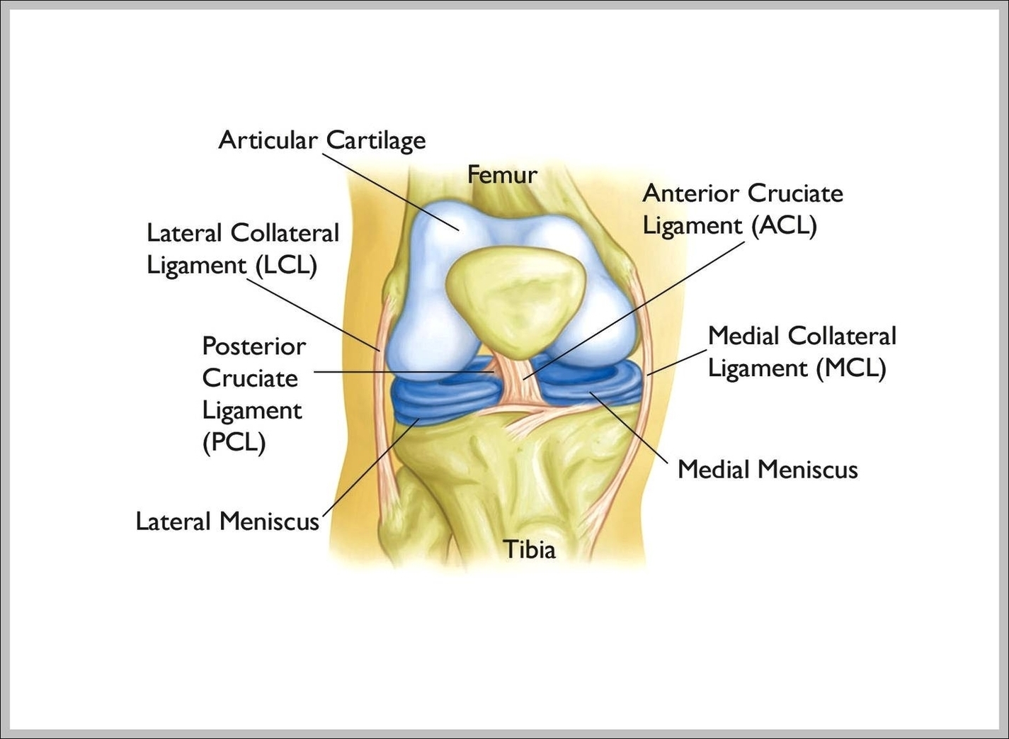
Knee Diagram Tendons Image
Knee joint is one of the most important hinge joints of our body. Its complexity and its efficiency is the best example of God’s creation. The anatomy of the knee consists of bones, muscles, nerves, cartilages, tendons and ligaments. All View Diagram Knee Diagram Tendons Image