T10 Vertebrae Image

This human anatomy diagram with labels depicts and explains the details and or parts of the T10 Vertebrae Image. Human anatomy diagrams and charts show internal organs, body systems, cells, conditions, sickness and symptoms information and/or tips to ensure one lives in good health.

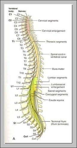
Also called the tenth thoracic vertebra, T10 is one of the twelve vertebrae of the thoracic spinal column. The nerves that control the muscles of the lower abdomen originate through here. What is the T11 Vertebra? The eleventh thoracic vertebra (T11) is one of the last thoracic spinal vertebrae.

The T1 vertebra is the first (uppermost) of the twelve (12) thoracic vertebrae that make up the central and largest section of the spinal column between the lumbar vertebrae below and the cervical vertebrae above. The second vertebra in the thoracic spine is responsible for helping to support the rib cage.

T9 (9th Thoracic Vertebra) The T9 vertebra is one of the lowest positioned of the twelve (12) thoracic vertebrae, and thus one of the largest. While the T2-T9 vertebrae are often linked descriptively, the T9 vertebra has certain particularities. Instead of two costal demi-facets (superior and inferior) on the exterior of the centrum, …

T10 Vertebrae Image

T10 Vertebrae Image


Tags: ,